What's new
  • As of today ICMag has his own Discord server. In this Discord server you can chat, talk with eachother, listen to music, share stories and pictures...and much more. Click the link to join now and let's grow together! https://discord.gg/2RRJW2XCZU
  • ICMag and The Vault are running a NEW contest in October! You can check it here. Prizes are seeds & forum premium access. Come join in!

... sig ...

acespicoli

Well-known member

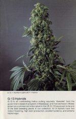
‘Thai Stick’ or ‘Lao Stick’?​

*
Bolikhamsai is one of the main production centres for export ganja right across the Mekong from northern Isan (Northeast Thailand) – both sides of the river being ethnic Lao and speaking Lao anywhere outside the towns.
*
In the ‘Thai Stick’ era, nominally ‘Thai’ ganja came from north Isan and Central Laos, one of the most famous batches being Central Lao ‘Golden Voice’ – as branded by Western smugglers…
*
Cultivation across north Isan was part of a very Lao tradition of growing and smoking ganja that expanded and commercialized with the major urban centre of the region, Vientiane.
*
In Vientiane’s decadent 60s Cold War heyday, the villagers of northern Isan supplied most of the young women who were trafficked or migrated to brothels, opium palaces, and go-go bars as the Lao economy boomed during America’s anti-Communist crusade in Indochina.
*
Western money also poured into provincial Isan towns such as Udon, now once again eclipsed by Vientiane, but then profiting from a commodity that could literally fetch more than its weight in gold in the USA.
*
The close careless planting in the photo is a sign of bulk production. But now that Thailand and Laos are eyeing full legalization, the crop is set to once again realize its potential and no doubt ultimately exceed the days when it was branded by Westerners as ‘Thai Stick’.

BBC JOURNALIST NEVER TRIED THAI GANJA, FEEDS ‘SKUNK’ HYSTERIA​

February 21, 2015 · by The Real Seed Company · in cannabis, CBD, drug policy, history, landraces, skunk. ·
David Shukman of the BBC claims that “The weed so familiar to many of my generation was characterised by a relatively balanced amount of THC and CBD” when compared to today’s hybrid strains. This is true only of traditional hashish from regions such as Afghanistan, Morocco, Lebanon and Nepal.
Because old-fashioned cannabis resin (‘hash’) is made from landraces which have been selected for resin production and had little selection for potency, it does indeed often exhibit something approximating a balanced 1:1 THC:CBD ratio. But even back in the ’60s and ’70s, an era in which we are told cannabis was a more innocent and far gentler plant, there was plenty of imported pot in which CBD was often more or less absent and THC levels could be very high. Typically, this took the form either of refined cannabis oil or—more usually—of high quality ganja, meaning cured unseeded or lightly seeded flowering tops from the tropics and subtropics – ‘Thai Stick’ and the like.
522
A traditional ganja strain flowering in Manipur, in the Indian tropics.
In Thailand, Malawi, or regions such as South India, the strains of cannabis that have been grown for centuries to produce ganja most often produce only minimal quantities of CBD and can even exhibit levels of THC comparable to those of so-called ‘skunk’. Back in the hippie heyday, such pot was given informal underground brands like ‘Swazi Red’ or ‘Colombian Gold’.
557
Traditional Indian ganja (‘herbal cannabis’) from Manipur, Northeast India.
In 1975 and 1976 the Laboratory of the Government Chemist reported Thai with as much as 17% THC—and that was after the bud had spent months on boats reaching England. THC levels such as this are far from unusual in Southeast Asia. In the ganja growing heartlands along the Mekong River generations of farmers have provided a continuous selective pressure for potency. Traditionally, a key part of the cannabis economy in Thailand and Laos is said to have been formed by specialists who produced high-quality seed, which was then supplied to farmers. The best of any season’s harvest would come from such fields. Similarly, in the Imphal Valley of Manipur, farmers and home growers know to keep seeds from good (i.e., potent) batches of ganja for sowing the following season. On an early collecting trip to Manipur, for example, I found a householder who only relucantly parted with seed from a standout plant. Growers who know this batch can attest to how potent the best individuals were.
566
A garden of ganja plants midway through harvest in Manipur, India.
Clearly the selective pressure in traditional ganja growing is less intensive than that of the modern so-called ‘clone’ method for breeding skunk, in which selected ‘mother plants’ can be kept indefinitely under 24-hour light regimes. However, the cumulative effect over generations of traditional ganja farming can result in very strong cannabis that’s often in any meaninful sense devoid of CBD. Analysis of samples from tropical India indicates that CBD is typically absent—even in some of the milder Bengali strains. This was the case in the ’60s and remains so today. Regarding THC levels, further evidence that the potency of modern hybrids is far from unprecedented comes, once again, from British seizures. In the words of the Home Office, as recorded in Hansard:
The latest data from the Forensic Science Service Ltd (FSS) show that the average tetrahydrocannabinol (THC) content of mature flowering tops from plants, otherwise known as sinsemilla, seized and submitted to the FSS from the 1 January 2008 to the present day was 14.0%. By comparison, during the same period, the average THC content of traditional imported cannabis and cannabis resin was 12.5% and 5.5% respectively.
On average, skunk (the FSS called it ‘sinsemilla’) showed only 1.5% more THC than ganja. So much for many times more potent. And, importantly, only in hashish would THC have been offset by a similar quantity of CBD.
In fairness, the BBC’s reporting on cannabis does seem to be improving. But—even if unwittingly—Aunty is still feeding the skunk hysteria.
Postscript:
Looking again at the LGC data I see that in ’78 a sample of Indian cannabis resin showed 26% THC. Customs bagged a 16% THC batch of Moroccan resin in ’75, and a shipment of Pakistani resin with the same strength in ’78. And then there are the concentrated extracts—‘hash oils’—with Indian, Kenyan and Pakistani samples all hitting around 40% THC. Simple extracts of this potency need to be made from starter material with a high cannabinoid content. The most potent was 48% THC oil from India seized in 1975. This is likely to have been prepared from tropical ganja plants rather than northern, Himalayan charas landraces.
The LGC does make the important point that “Cannabis resins normally had higher THC contents than most herbal material…”. Typically, the ‘herbal cannabis’ reaching UK shores during this era was of fairly low potency—mid to low single figures THC—either because it was from poor stock, or had degraded en route. This does, to be fair, support the kind of line Shukman is taking, though not the current media focus on CBD. Historically, ganja is most unlikely to have contained CBD in any relevant quantity. Some of the best bud to reach Britain in the ’70s appears to have been coming from southern Africa, such as Rhodesia—now Zimbabwe—with a sample hitting 12%. Nigerian and South Indian ganja seizures were milder, at 7.4% and 7.8%—less potent, but still typically without any meaningful quantity of CBD.
Manipuri (2)

Manipuri ganja landrace in full flower, Imphal Valley, Northeast India
Should Thai ganja be seen as exceptional? Perhaps for this era in the UK it should, though both the proportion of samples available from seizures and ample anecdotal evidence suggest there was plenty of it about in the country, in addition to the stronger resins and concentrated oils. The LGC points out the unique appearance of the “Thai Stick” brand: “Green or brown sticks of several seedless tops tied around bamboo with a number of sticks compressed into a slab.” I’m sure there are readers who recognise this, either from having seen the real thing back in the day or on their travels.
40960929_2235918826450616_195626865455529984_n

Thai sticks, as shared on Facebook by Peter Maguire, author of Thai Stick: Surfers, Scammers, and the Untold Story of the Marijuana Trade
The LGC figures suggest that the potency of Thai seizures peaked in the early to mid ’70s, and then began to decline after the Vietnam War and the Fall of Saigon. It’s not clear whether this sample is indicative of changes at the source in Isan, Northeast Thailand. But as the authors state, in ’75 and ’76 “by far the highest quality cannabis originated in South East Asia (exclusively in the form of “Thai sticks”) and this was reflected in its street price, at least in the United Kingdom, over the same period. However, the 1978 seizures which originated in Thailand, while still prepared in the form of sticks, showed a dramatic decrease in THC level compared with previous years. A careful study of the physical appearance of seizures of Thai origin for the three years revealed an increasing seed content in the cannabis.”
To take a contrarian line, perhaps the changes that have occurred in the UK over the last two decades are about catching up to highs the market, and its consumers, had already reached in 1975.
The featured image on the homepage is via the authors of a superb new book, Thai Stick: Surfers, Scammers, and the Untold Story of the International Marijuana Trade.
Mango Thai crop2

PC*
 
Last edited:
  • Like
Reactions: Rgd

acespicoli

Well-known member
Logo The Seed Bank


Northern Lights #2 is a mostly indica variety from The Seed Bank and can be cultivated indoors. The Seed Banks Northern Lights #2 is a THC dominant variety and is/was never available as feminized seeds.
sturdy, pine tree shaped plant
odor: musty and piney


»»» Purest Indica x Afghani by Don the Indian

The Seed Banks Hash Plant / Northern Lights #1 F-1 Hybrid Description​


Logo The Seed Bank Hash Plant is named for its hashy-tasting, highly resinous buds. One of the most famous indoor cuttings from the Northwest USA, Hash Plant / Northern Lights #1 F-1 Hybrid has produced some of the highest quality F-1 Hybrids that we have seen, High potency, abundant resin, and an extremely fast finish, with very strong Indica high, sophisticated taste and aroma. Excellent indoor plant for personal stash or select commercial trade. Here in Holland, even experienced smokers have had trouble finishing a Joint of Hash Plant / Northern Lights # 1. It has certainly produced the strongest specimens that I have ever smoked, with a very pleasant, narcotic-type high. We are now selecting for the best NL #1 males to use in this tremendous hybrid.

Indoor height at 100 days: 36-50 inches.
Indoor yield at 100 days: up to 85 grams.
Indoor flowering period at 12 hrs. darkness: 40-45 days.
Hash Plant / Northern Lights #1 F-1 Hybrid

The Seed Banks G-13 x Hash Plant F-1 Hybrid Description​


Logo The Seed Bank One of the fastest-flowering indoor hybrids that we have yet produced. It has the marvellous potency and quick finish of Hash Plant, plus vigor and yield. In our lest test, all seedlings finished in less than 50 days of flowering, producing beautifully solid, heavy buds. Clones from the seedlings rooted very easily and should flower even more quickly. Powerful all-Indica high, but not brutal. This hybrid has perhaps the best balance of speed, yield and quality, destined for commercial success!

Indoor height at 100 days: 36-50 inches.
Indoor yield at 100 days: up to 100 grams.
Indoor flowering period at 12 hrs. darkness: 45-50 days.


Neville (The Seed Bank) write in his 1987er-catalouge:

G-13 is an outstanding indica cutting reputedly 'liberated' from the government research program in Mississippi, and now we have it. Widely grown as commercial indoor plant in the US, G-13 has proven to be one of the best breeding plants in our collection. G-13 hybrids have fast indoor flowering, high resin production, excellent potency, and extreme hybrid vigor.

DPH Orig Drop 88G13HP.png

screenshot-www_icmag_com-2024_10_09-08_35_04.png
 

Attachments

  • SeedBank88_3_no3.jpg
    SeedBank88_3_no3.jpg
    51.7 KB · Views: 9
  • SeedBank88_no2.jpg
    SeedBank88_no2.jpg
    43.3 KB · Views: 9
  • SeedBank88_1.jpg
    SeedBank88_1.jpg
    40 KB · Views: 5
  • screenshot-mrnice_nl-2024_10_09-08_32_22.png
    screenshot-mrnice_nl-2024_10_09-08_32_22.png
    176.5 KB · Views: 4
Last edited:

acespicoli

Well-known member
Icon image Field Book
Field Book
4.5star

Icon image Prospector
Prospector

Icon image Intercross
Intercross

Icon image Verify
Verify
3.1star

Icon image OneKK
OneKK

Icon image Coordinate
Coordinate

Role in evolution

By introducing novel genetic qualities to a population of organisms, de novo mutations play a critical role in the combined forces of evolutionary change. However, the weight of genetic diversity generated by mutational change is often considered a generally "weak" evolutionary force.[102] Although the random emergence of mutations alone provides the basis for genetic variation across all organic life, this force must be taken in consideration alongside all evolutionary forces at play. Spontaneous de novo mutations as cataclysmic events of speciation depend on factors introduced by natural selection, genetic flow, and genetic drift. For example, smaller populations with heavy mutational input (high rates of mutation) are prone to increases of genetic variation which lead to speciation in future generations. In contrast, larger populations tend to see lesser effects of newly introduced mutated traits. In these conditions, selective forces diminish the frequency of mutated alleles, which are most often deleterious, over time.[119]

Monohybrid cross​

Main article: Monohybrid cross
"Mono-" means "one"; this cross indicates that the examination of a single trait. This could mean (for example) eye color. Each genetic locus is always represented by two letters. So in the case of eye color, say "B = Brown eyes" and "b = green eyes". In this example, both parents have the genotype Bb. For the example of eye color, this would mean they both have brown eyes. They can produce gametes that contain either the B or the b allele. (It is conventional in genetics to use capital letters to indicate dominant alleles and lower-case letters to indicate recessive alleles.) The probability of an individual offspring's having the genotype BB is 25%, Bb is 50%, and bb is 25%. The ratio of the phenotypes is 3:1, typical for a monohybrid cross. When assessing phenotype from this, "3" of the offspring have "Brown" eyes and only one offspring has "green" eyes. (3 are "B?" and 1 is "bb")

Paternal

Maternal
Bb
BBBBb
bBbbb
The way in which the B and b alleles interact with each other to affect the appearance of the offspring depends on how the gene products (proteins) interact (see Mendelian inheritance). This can include lethal effects and epistasis (where one allele masks another, regardless of dominant or recessive status).

Dihybrid cross​

Main article: Dihybrid cross
More complicated crosses can be made by looking at two or more genes. The Punnett square works, however, only if the genes are independent of each other, which means that having a particular allele of gene "A" does not alter the probability of possessing an allele of gene "B". This is equivalent to stating that the genes are not linked, so that the two genes do not tend to sort together during meiosis.

The following example illustrates a dihybrid cross between two double-heterozygote pea plants. R represents the dominant allele for shape (round), while r represents the recessive allele (wrinkled). A represents the dominant allele for color (yellow), while a represents the recessive allele (green). If each plant has the genotype RrAa, and since the alleles for shape and color genes are independent, then they can produce four types of gametes with all possible combinations: RA, Ra, rA, and ra.

RARarAra
RARRAARRAaRrAARrAa
RaRRAaRRaaRrAaRraa
rARrAARrAarrAArrAa
raRrAaRraarrAarraa
Since dominant traits mask recessive traits (assuming no epistasis), there are nine combinations that have the phenotype round yellow, three that are round green, three that are wrinkled yellow, and one that is wrinkled green. The ratio 9:3:3:1 is the expected outcome when crossing two double-heterozygous parents with unlinked genes. Any other ratio indicates that something else has occurred (such as lethal alleles, epistasis, linked genes, etc.).

Forked-line method​

The forked-line method (also known as the tree method and the branching system) can also solve dihybrid and multi-hybrid crosses. A problem is converted to a series of monohybrid crosses, and the results are combined in a tree. However, a tree produces the same result as a Punnett square in less time and with more clarity. The example below assesses another double-heterozygote cross using RrYy x RrYy. As stated above, the phenotypic ratio is expected to be 9:3:3:1 if crossing unlinked genes from two double-heterozygotes. The genotypic ratio was obtained in the diagram below, this diagram will have more branches than if only analyzing for phenotypic ratio.


Importance to evolutionary biology​

According to Lewontin,[5] the theoretical task for population genetics is a process in two spaces: a "genotypic space" and a "phenotypic space". The challenge of a complete theory of population genetics is to provide a set of laws that predictably map a population of genotypes (G1) to a phenotype space (P1), where selection takes place, and another set of laws that map the resulting population (P2) back to genotype space (G2) where Mendelian genetics can predict the next generation of genotypes, thus completing the cycle. Even if non-Mendelian aspects of molecular genetics are ignored, this is a gargantuan task. Visualizing the transformation schematically:

{\displaystyle G_{1}\;{\stackrel {T_{1}}{\rightarrow }}\;P_{1}\;{\stackrel {T_{2}}{\rightarrow }}\;P_{2}\;{\stackrel {T_{3}}{\rightarrow }}\;G_{2}\;{\stackrel {T_{4}}{\rightarrow }}\;G_{1}'\;\rightarrow \cdots }

(adapted from Lewontin 1974, p. 12). T1 represents the genetic and epigenetic laws, the aspects of functional biology, or development, that transform a genotype into phenotype. This is the "genotype–phenotype map". T2 is the transformation due to natural selection, T3 are epigenetic relations that predict genotypes based on the selected phenotypes and finally T4 the rules of Mendelian genetics.

In practice, there are two bodies of evolutionary theory that exist in parallel, traditional population genetics operating in the genotype space and the biometric theory used in plant and animal breeding, operating in phenotype space. The missing part is the mapping between the genotype and phenotype space. This leads to a "sleight of hand" (as Lewontin terms it) whereby variables in the equations of one domain, are considered parameters or constants, where, in a full-treatment, they would be transformed themselves by the evolutionary process and are functions of the state variables in the other domain. The "sleight of hand" is assuming that the mapping is known. Proceeding as if it is understood is enough to analyze many cases of interest. For example, if the phenotype is almost one-to-one with genotype (sickle-cell disease) or the time-scale is sufficiently short, the "constants" can be treated as such; however, there are also many situations where that assumption does not hold.


Genotype–phenotype map​


Genotype-Phenotype Map
A very simple genotype–phenotype map that only shows additive pleiotropy effects.
The genotype–phenotype map is a conceptual model in genetic architecture. Coined in a 1991 paper by Pere Alberch,[1] it models the interdependency of genotype (an organism's full hereditary information) with phenotype (an organism's actual observed properties).

The map visualises a relationship between genotype & phenotype which, crucially:[2]

  1. is of greater complexity than a straightforward one-to-one mapping of genotype to/from phenotype.
  2. accommodates a parameter space, along which at different points a given phenotype is said to be more or less stable.
  3. accommodates transformational boundaries in the parameter space, which divide phenotype states from one another.
  4. accounts for different polymorphism and/or polyphenism in populations, depending on their area of parameter space they occupy.
Evolvability is literally defined as the ability to evolve. In terms of genetics, evolvability is the ability of a genetic system to produce and maintain potentially adaptive genetic variants. There are several aspects of genetic architecture that contribute strongly to the evolvability of a system, including autonomy, mutability, coordination, epistasis, pleiotropy, polygeny, and robustness.[1][2]

  • Autonomy: the existence of quasi-independent characters with the potential for evolutionary autonomy.[5]
  • Mutability: the possibility that genetic mutation can occur.
  • Coordination: a phenomenon such as development, during which many different genetic processes and changes happen at once.
  • Epistasis: a phenomenon in which one gene is dependent on the presence of one or more "modifier" genes.
  • Polygeny: a phenomenon in which multiple genes contribute to a particular phenotypic character.
  • Pleiotropy: a phenomenon in which a single gene affects one or more phenotypic characteristics.
  • Robustness: the ability of a phenotype to remain constant in spite of genetic mutation.

Diploid organisms​

Epistasis in diploid organisms is further complicated by the presence of two copies of each gene. Epistasis can occur between loci, but additionally, interactions can occur between the two copies of each locus in heterozygotes. For a two locus, two allele system, there are eight independent types of gene interaction.

Additive A locusAdditive B locusDominance A locusDominance B locus
aaaAAAaaaAAAaaaAAAaaaAAA
bb10–1bb111bb–11–1bb–1–1–1
bB10–1bB000bB–11–1bB111
BB10–1BB–1–1–1BB–11–1BB–1–1–1
Additive by Additive EpistasisAdditive by Dominance EpistasisDominance by Additive EpistasisDominance by Dominance Epistasis
aaaAAAaaaAAAaaaAAAaaaAAA
bb10–1bb10–1bb1–11bb–11–1
bB000bB–101bB000bB1–11
BB–101BB10–1BB–11–1BB–11–1
1728528015236.png

The schematic, screenshots of representative resources and a user case of CannabisGDB. (a) The flow diagram showing design and construction of CannabisGDB. (b)The home page of CannabisGDB. (c)The ‘varieties module’ providing summary of cannabis genomes, detailed information of cannabis varieties and genome browser tool. (d) The ‘gene loci module’ showing detailed information of genes identified in this study. (e) The ‘metabolites module’ providing chemical phenotypes in various cannabis varieties. (f) The ‘proteins module’ presenting information of experimentally identified proteins. (g) A case study for the application of CannabisGDB. Dashed lines indicate linkages between different pages.
doi: 10.1111/pbi.13548


A​

B​

C​

D​

E​

F​

G​

H​

I​

K​

M​

N​

O​

P​

R​

S​

Categories:
Hidden category:

Post needs some cleaning 🛌
 
Last edited:

acespicoli

Well-known member
The history of scientific method considers changes in the methodology of scientific inquiry, as distinct from the history of science itself. The development of rules for scientific reasoning has not been straightforward; scientific method has been the subject of intense and recurring debate throughout the history of science, and eminent natural philosophers and scientists have argued for the primacy of one or another approach to establishing scientific knowledge.

This post deserves its own thread?

The philosopher Wesley C. Salmon described scientific inquiry:

The search for scientific knowledge ends far back into antiquity. At some point in the past, at least by the time of Aristotle, philosophers recognized that a fundamental distinction should be drawn between two kinds of scientific knowledge—roughly, knowledge that and knowledge why. It is one thing to know that each planet periodically reverses the direction of its motion with respect to the background of fixed stars; it is quite a different matter to know why. Knowledge of the former type is descriptive; knowledge of the latter type is explanatory. It is explanatory knowledge that provides scientific understanding of the world. (Salmon, 2006, pg. 3)[1]
One way of describing scientific method would then contain these steps as a minimum:

  1. Make a set of observations regarding the phenomenon being studied.
  2. Form a hypothesis that might explain the observations. (This may involve inductive and/or abductive reasoning.)
  3. Identify the implications and outcomes that must follow, if the hypothesis is to be true.
  4. Perform other experiments or observations to see if any of the predicted outcomes fail.
  5. If any predicted outcomes fail, the hypothesis is proven false since if A implies B, then not B implies not A (by deduction). It is then necessary to change the hypothesis and go back to step 3. If the predicted outcomes are confirmed, the hypothesis is not proved, but rather can be said to be consistent with known data.
When a hypothesis has survived a sufficient number of tests, it may be promoted to a scientific theory. A theory is a hypothesis that has survived many tests and seems to be consistent with other established scientific theories. Since a theory is a promoted hypothesis, it is of the same 'logical' species and shares the same logical limitations. Just as a hypothesis cannot be proven but can be disproved, that same is true for a theory. It is a difference of degree, not kind.
1728529032536.png
 
Last edited:

acespicoli

Well-known member

Plants​


Phenotypic plasticity of sorghum flowering time evaluated from seven environments. The identified photothermal time, a performance-independent index, quantifies the relevant environmental input and enables a systematic framework for modelling, explaining, and predicting phenotypic values under natural conditions.[6]
Phenotypic plasticity in plants includes the timing of transition from vegetative to reproductive growth stage, the allocation of more resources to the roots in soils that contain low concentrations of nutrients, the size of the seeds an individual produces depending on the environment,[7] and the alteration of leaf shape, size, and thickness.[8] Leaves are particularly plastic, and their growth may be altered by light levels. Leaves grown in the light tend to be thicker, which maximizes photosynthesis in direct light; and have a smaller area, which cools the leaf more rapidly (due to a thinner boundary layer). Conversely, leaves grown in the shade tend to be thinner, with a greater surface area to capture more of the limited light.[9][10] Dandelion are well known for exhibiting considerable plasticity in form when growing in sunny versus shaded environments. The transport proteins present in roots also change depending on the concentration of the nutrient and the salinity of the soil.[11] Some plants, Mesembryanthemum crystallinum for example, are able to alter their photosynthetic pathways to use less water when they become water- or salt-stressed.[12]

Because of phenotypic plasticity, it is hard to explain and predict the traits when plants are grown in natural conditions unless an explicit environment index can be obtained to quantify environments. Identification of such explicit environment indices from critical growth periods being highly correlated with sorghum and rice flowering time enables such predictions.[6][13] Additional work is being done to support the agricultural industry, which faces severe challenges in prediction of crop phenotypic expression in changing environments. Since many crops supporting the global food supply are grown in a wide variety of environments, understanding and ability to predict crop genotype by environment interaction will be essential for future food stability.[14]

Phytohormones and leaf plasticity​

Leaves are very important to a plant in that they create an avenue where photosynthesis and thermoregulation can occur. Evolutionarily, the environmental contribution to leaf shape allowed for a myriad of different types of leaves to be created.[15] Leaf shape can be determined by both genetics and the environment.[16] Environmental factors, such as light and humidity, have been shown to affect leaf morphology,[17] giving rise to the question of how this shape change is controlled at the molecular level. This means that different leaves could have the same gene but present a different form based on environmental factors. Plants are sessile, so this phenotypic plasticity allows the plant to take in information from its environment and respond without changing its location.

In order to understand how leaf morphology works, the anatomy of a leaf must be understood. The main part of the leaf, the blade or lamina, consists of the epidermis, mesophyll, and vascular tissue. The epidermis contains stomata which allows for gas exchange and controls perspiration of the plant. The mesophyll contains most of the chloroplast where photosynthesis can occur. Developing a wide blade/lamina can maximize the amount of light hitting the leaf, thereby increasing photosynthesis, however too much sunlight can damage the plant. Wide lamina can also catch wind easily which can cause stress to the plant, so finding a happy medium is imperative to the plants’ fitness. The Genetic Regulatory Network is responsible for creating this phenotypic plasticity and involves a variety of genes and proteins regulating leaf morphology. Phytohormones have been shown to play a key role in signaling throughout the plant, and changes in concentration of the phytohormones can cause a change in development.[18]

Studies on the aquatic plant species Ludwigia arcuata have been done to look at the role of abscisic acid (ABA), as L. arcuata is known to exhibit phenotypic plasticity and has two different types of leaves, the aerial type (leaves that touch the air) and the submerged type (leaves that are underwater).[19] When adding ABA to the underwater shoots of L. arcuata, the plant was able to produce aerial type leaves underwater, suggesting that increased concentrations of ABA in the shoots, likely caused by air contact or a lack of water, triggers the change from the submerged type of leaf to the aerial type. This suggests ABA's role in leaf phenotypic change and its importance in regulating stress through environmental change (such as adapting from being underwater to above water). In the same study, another phytohormone, ethylene, was shown to induce the submerged leaf phenotype unlike ABA, which induced aerial leaf phenotype. Because ethylene is a gas, it tends to stay endogenously within the plant when underwater – this growth in concentration of ethylene induces a change from aerial to submerged leaves and has also been shown to inhibit ABA production, further increasing the growth of submerged type leaves. These factors (temperature, water availability, and phytohormones) contribute to changes in leaf morphology throughout a plants lifetime and are vital to maximize plant fitness.
 

acespicoli

Well-known member

21 strains found​

Type I Plants
THC + THCA %20+ Reverse sort order
  1. Red Eye OG

    CCLV
    Jun 26, 2019
    THC + THCA: 56.4% (Moon Rocks?)
  2. Headcheese

    Polaris Wellness Center
    Jun 26, 2019
    THC + THCA: 31.973%
  3. Mothers Milk #5

    Slater Center
    Jun 26, 2019
    THC + THCA: 30.59%
  4. Blue Dream

    QualCan
    Sep 17, 2017
    THC + THCA: 28.273%
  5. Miss X

    DigiPath Labs
    Aug 28, 2017
    THC + THCA: 27%
  6. East Coast Sour Diesel

    Flower Power Botanicals LLC
    May 22, 2016
    THC + THCA: 26.872%
  7. Blue Dream

    CW NV Cultivators
    Sep 17, 2017
    THC + THCA: 26.64%
  8. Durban Poison

    DigiPath Labs
    Sep 17, 2017
    THC + THCA: 25.7%
  9. Golden Goat 2

    DigiPath Labs
    Aug 27, 2017
    THC + THCA: 25.7%
  10. Blueberry Cheesecake

    Practical Possiblities
    Feb 20, 2017
    THC + THCA: 25.5%
  11. Blue Dream

    CW NV Cultivators
    Sep 17, 2017
    THC + THCA: 25.333%
  12. Skywalker OG

    THC Design
    Mar 28, 2017
    THC + THCA: 25.06%
  13. Durban Poison #1

    DigiPath Labs
    Sep 17, 2017
    THC + THCA: 24.9%
  14. Durban Poison #1

    DigiPath Labs
    Aug 28, 2017
    THC + THCA: 24.9%
  15. Chem 91

    Slater Center
    Jun 26, 2019
    THC + THCA: 23.61%
  16. Durban Poison

    DigiPath Labs
    Aug 28, 2017
    THC + THCA: 23.3%
  17. Kimbo Slice

    DigiPath Labs
    Aug 28, 2017
    THC + THCA: 23.1%
  18. Blue Dream

    Silver Sage Wellness
    Sep 17, 2017
    THC + THCA: 22.734%
  19. Blue Dream

    Flora Vega
    Sep 17, 2017
    THC + THCA: 21.369%
  20. Snoops Dream

    Avitas
    Nov 19, 2017
    THC + THCA: 20.4%
  21. Snoops Dream

    Avitas
    Sep 17, 2017
    THC + THCA: 20.4%
 

acespicoli

Well-known member
The Emerald Triangle is a region in Northern California that derives its name from being the largest cannabis-producing region in the United States. The region includes three counties in an upside-down triangular configuration:

screenshot-en_wikipedia_org-2024_10_10-16_15_26.png

flowering period of 49–63 days.

Properties and Effects of Gary Payton​


The effects of Gary Payton can vary depending on several variables, including light, stress, and nutrients. However, this strain typically delivers a potent and fast-acting high defined by giggles, euphoria, and sociability. Tests show that THC levels usually average around 23%. This places Gary Payton in the middle ground between weaker old-school varieties and some of the most potent modern cultivars. With that said, new smokers should approach these buds sensibly and slowly.


While THC creates the core effects of this strain, several other phytochemicals also contribute to the experience. Gary Payton buds produce various aromatic terpenes that contribute to the aroma, taste, and overall effects of the strains. Among these compounds, caryophyllene, limonene, and pinene are the most dominant.


Caryophyllene plays a similar role to a cannabinoid, binding to CB2 receptors in the body. Put simply, this produces a physically soothing effect that can help to reduce physical discomfort. Limonene adds stimulation to the equation and underpins the energizing impact of Gary Payton. Finally, pinene clears the mind and helps to keep the short-term memory impairment of THC at bay.


Uses of Gary Payton​


Both recreational and medical users report several key uses of this strain. You'll find that Gary Payton quickly induces the munchies. This property makes the strain a good candidate for stimulating the appetite of medical users struggling with the thought of getting food down.


Many users also report that Gary Payton makes them feel giggly and talkative. These buds ramp up dopamine levels in the brain, which makes this strain attractive to those looking for respite from low mood. Moreover, terpenes such as limonene and pinene show promise when it comes to relieving mood-related disorders. Because this strain contains high levels of these molecules, it helps smokers to gain confidence at the start of the day and wind down in the evenings.


Aside from the medicinal side of things, Gary Payton also helps to bolster creativity and divergent thinking in some users. Blazing up a large bong bowl or blunt before sitting down to write music or paint helps to get the creative cogs turning. If you're somebody that likes to use cannabis before hiking or hitting the gym, you'll find this strain can increase focus and alertness. Most fittingly, it also helps with concentration during casual hoop-shooting sessions!

OG Kush South Florida
 

acespicoli

Well-known member
Quantum biology

is the study of applications of quantum mechanics and theoretical chemistry to aspects of biology that cannot be accurately described by the classical laws of physics.[1] An understanding of fundamental quantum interactions is important because they determine the properties of the next level of organization in biological systems.

Many biological processes involve the conversion of energy into forms that are usable for chemical transformations, and are quantum mechanical in nature. Such processes involve chemical reactions, light absorption, formation of excited electronic states, transfer of excitation energy, and the transfer of electrons and protons (hydrogen ions) in chemical processes, such as photosynthesis, olfaction and cellular respiration.[2] Moreover, quantum biology may use computations to model biological interactions in light of quantum mechanical effects.[3] Quantum biology is concerned with the influence of non-trivial quantum phenomena,[4] which can be explained by reducing the biological process to fundamental physics, although these effects are difficult to study and can be speculative.[5]

Currently, there exist four major life processes that have been identified as influenced by quantum effects: enzyme catalysis, sensory processes, energy transference, and information encoding.[6]

History​

Quantum biology is an emerging field, in the sense that most current research is theoretical and subject to questions that require further experimentation. Though the field has only recently received an influx of attention, it has been conceptualized by physicists throughout the 20th century. It has been suggested that quantum biology might play a critical role in the future of the medical world.[7] Early pioneers of quantum physics saw applications of quantum mechanics in biological problems. Erwin Schrödinger's 1944 book What Is Life? discussed applications of quantum mechanics in biology.[8] Schrödinger introduced the idea of an "aperiodic crystal" that contained genetic information in its configuration of covalent chemical bonds. He further suggested that mutations are introduced by "quantum leaps". Other pioneers Niels Bohr, Pascual Jordan, and Max Delbrück argued that the quantum idea of complementarity was fundamental to the life sciences.[9] In 1963, Per-Olov Löwdin published proton tunneling as another mechanism for DNA mutation. In his paper, he stated that there is a new field of study called "quantum biology".[10] In 1979, the Soviet and Ukrainian physicist Alexander Davydov published the first textbook on quantum biology entitled Biology and Quantum Mechanics.[11][12]

Enzyme catalysis​

Enzymes have been postulated to use quantum tunneling to transfer electrons in electron transport chains.[13][14][15] It is possible that protein quaternary architectures may have adapted to enable sustained quantum entanglement and coherence, which are two of the limiting factors for quantum tunneling in biological entities.[16] These architectures might account for a greater percentage of quantum energy transfer, which occurs through electron transport and proton tunneling (usually in the form of hydrogen ions, H+).[17][18] Tunneling refers to the ability of a subatomic particle to travel through potential energy barriers.[19] This ability is due, in part, to the principle of complementarity, which holds that certain substances have pairs of properties that cannot be measured separately without changing the outcome of measurement. Particles, such as electrons and protons, have wave-particle duality; they can pass through energy barriers due to their wave characteristics without violating the laws of physics. In order to quantify how quantum tunneling is used in many enzymatic activities, many biophysicists utilize the observation of hydrogen ions. When hydrogen ions are transferred, this is seen as a staple in an organelle's primary energy processing network; in other words, quantum effects are most usually at work in proton distribution sites at distances on the order of an angstrom (1 Å).[20][21] In physics, a semiclassical (SC) approach is most useful in defining this process because of the transfer from quantum elements (e.g. particles) to macroscopic phenomena (e.g. biochemicals). Aside from hydrogen tunneling, studies also show that electron transfer between redox centers through quantum tunneling plays an important role in enzymatic activity of photosynthesis and cellular respiration (see also Mitochondria section below).[15][22]

Ferritin​

Ferritin is an iron storage protein that is found in plants and animals. It is usually formed from 24 subunits that self-assemble into a spherical shell that is approximately 2 nm thick, with an outer diameter that varies with iron loading up to about 16 nm. Up to ~4500 iron atoms can be stored inside the core of the shell in the Fe3+ oxidation state as water-insoluble compounds such as ferrihydrite and magnetite.[23] Ferritin is able to store electrons for at least several hours, which reduce the Fe3+ to water soluble Fe2+.[24] Electron tunneling as the mechanism by which electrons transit the 2 nm thick protein shell was proposed as early as 1988.[25] Electron tunneling and other quantum mechanical properties of ferritin were observed in 1992,[26] and electron tunneling at room temperature and ambient conditions was observed in 2005.[27] Electron tunneling associated with ferritin is a quantum biological process, and ferritin is a quantum biological agent.

Electron tunneling through ferritin between electrodes is independent of temperature, which indicates that it is substantially coherent and activation-less.[28] The electron tunneling distance is a function of the size of the ferritin. Single electron tunneling events can occur over distances of up to 8 nm through the ferritin, and sequential electron tunneling can occur up to 12 nm through the ferritin. It has been proposed that the electron tunneling is magnon-assisted and associated with magnetite microdomains in the ferritin core.[29]

Early evidence of quantum mechanical properties exhibited by ferritin in vivo was reported in 2004, where increased magnetic ordering of ferritin structures in placental macrophages was observed using small angle neutron scattering (SANS).[30] Quantum dot solids also show increased magnetic ordering in SANS testing,[31] and can conduct electrons over long distances.[32] Increased magnetic ordering of ferritin cores disposed in an ordered layer on a silicon substrate with SANS testing has also been observed.[33] Ferritin structures like those in placental macrophages have been tested in solid state configurations and exhibit quantum dot solid-like properties of conducting electrons over distances of up to 80 microns through sequential tunneling and formation of Coulomb blockades.[34][35][36] Electron transport through ferritin in placental macrophages may be associated with an anti-inflammatory function.[37]

Conductive atomic force microscopy of substantia nigra pars compacta (SNc) tissue demonstrated evidence of electron tunneling between ferritin cores, in structures that correlate to layers of ferritin outside of neuromelanin organelles.[38]

Evidence of ferritin layers in cell bodies of large dopamine neurons of the SNc and between those cell bodies in glial cells has also been found,[39][40][41] and is hypothesized to be associated with neuron function.[42] Overexpression of ferritin reduces the accumulation of reactive oxygen species (ROS),[43] and may act as a catalyst by increasing the ability of electrons from antioxidants to neutralize ROS through electron tunneling. Ferritin has also been observed in ordered configurations in lysosomes associated with erythropoiesis,[44] where it may be associated with red blood cell production. While direct evidence of tunneling associated with ferritin in vivo in live cells has not yet been obtained, it may be possible to do so using QDs tagged with anti-ferritin, which should emit photons if electrons stored in the ferritin core tunnel to the QD.[45]

  • Electron microscope image of placental macrophage ferritin
    Electron microscope image of placental macrophage ferritin
  • Conductive atomic force microscopy image of human substantia nigra pars compacta (SNc) tissue
    Conductive atomic force microscopy image of human substantia nigra pars compacta (SNc) tissue
  • Electron spectroscopic imaging of iron (red) outside of neuromelanin organelles
    Electron spectroscopic imaging of iron (red) outside of neuromelanin organelles
  • Electron microscope image of glial cell from SNc showing structures similar to ferritin in placental tissue
    Electron microscope image of glial cell from SNc showing structures similar to ferritin in placental tissue

Sensory processes​

[edit]

Olfaction​

Olfaction, the sense of smell, can be broken down into two parts; the reception and detection of a chemical, and how that detection is sent to and processed by the brain. This process of detecting an odorant is still under question. One theory named the "shape theory of olfaction" suggests that certain olfactory receptors are triggered by certain shapes of chemicals and those receptors send a specific message to the brain.[46] Another theory (based on quantum phenomena) suggests that the olfactory receptors detect the vibration of the molecules that reach them and the "smell" is due to different vibrational frequencies, this theory is aptly called the "vibration theory of olfaction."

The vibration theory of olfaction, created in 1938 by Malcolm Dyson[47] but reinvigorated by Luca Turin in 1996,[48] proposes that the mechanism for the sense of smell is due to G-protein receptors that detect molecular vibrations due to inelastic electron tunneling, tunneling where the electron loses energy, across molecules.[48] In this process a molecule would fill a binding site with a G-protein receptor. After the binding of the chemical to the receptor, the chemical would then act as a bridge allowing for the electron to be transferred through the protein. As the electron transfers across what would otherwise have been a barrier, it loses energy due to the vibration of the newly-bound molecule to the receptor. This results in the ability to smell the molecule.[48][4]

While the vibration theory has some experimental proof of concept,[49][50] there have been multiple controversial results in experiments. In some experiments, animals are able to distinguish smells between molecules of different frequencies and same structure,[51] while other experiments show that people are unaware of distinguishing smells due to distinct molecular frequencies.[52]

Vision​

Main article: Visual phototransduction
Vision relies on quantized energy in order to convert light signals to an action potential in a process called phototransduction. In phototransduction, a photon interacts with a chromophore in a light receptor. The chromophore absorbs the photon and undergoes photoisomerization. This change in structure induces a change in the structure of the photo receptor and resulting signal transduction pathways lead to a visual signal. However, the photoisomerization reaction occurs at a rapid rate, in under 200 femtoseconds,[53] with high yield. Models suggest the use of quantum effects in shaping the ground state and excited state potentials in order to achieve this efficiency.[54]

The sensor in the retina of the human eye is sensitive enough to detect a single photon.[55] Single photon detection could lead to multiple different technologies. One area of development is in quantum communication and cryptography. The idea is to use a biometric system to measure the eye using only a small number of points across the retina with random flashes of photons that "read" the retina and identify the individual.[56] This biometric system would only allow a certain individual with a specific retinal map to decode the message. This message can not be decoded by anyone else unless the eavesdropper were to guess the proper map or could read the retina of the intended recipient of the message.[57]

Energy transfer​

[edit]

Photosynthesis​

Main article: Photosynthesis
Generic photosystem Complex Antennae complex found in photosystems of both prokaryotes and eukaryotes Diagram of FMO complex. Light excites electrons in an antenna. The excitation then transfers through various proteins in the FMO complex to the reaction center to further photosynthesis.
Photosynthesis refers to the biological process that photosynthetic cells use to synthesize organic compounds from inorganic starting materials using sunlight.[58] What has been primarily implicated as exhibiting non-trivial quantum behaviors is the light reaction stage of photosynthesis. In this stage, photons are absorbed by the membrane-bound photosystems. Photosystems contain two major domains, the light-harvesting complex (antennae) and the reaction center. These antennae vary among organisms. For example, bacteria use circular aggregates of chlorophyll pigments, while plants use membrane-embedded protein and chlorophyll complexes.[59][60] Regardless, photons are first captured by the antennae and passed on to the reaction-center complex. Various pigment-protein complexes, such as the FMO complex in green sulfur bacteria, are responsible for transferring energy from antennae to reaction site. The photon-driven excitation of the reaction-center complex mediates the oxidation and the reduction of the primary electron acceptor, a component of the reaction-center complex. Much like the electron transport chain of the mitochondria, a linear series of oxidations and reductions drives proton (H+) pumping across the thylakoid membrane, the development of a proton motive force, and energetic coupling to the synthesis of ATP.

Previous understandings of electron-excitation transference (EET) from light-harvesting antennae to the reaction center have relied on the Förster theory of incoherent EET, postulating weak electron coupling between chromophores and incoherent hopping from one to another. This theory has largely been disproven by FT electron spectroscopy experiments that show electron absorption and transfer with an efficiency of above 99%,[61] which cannot be explained by classical mechanical models. Instead, as early as 1938, scientists theorized that quantum coherence was the mechanism for excitation-energy transfer. Indeed, the structure and nature of the photosystem places it in the quantum realm, with EET ranging from the femto- to nanosecond scale, covering sub-nanometer to nanometer distances.[62] The effects of quantum coherence on EET in photosynthesis are best understood through state and process coherence. State coherence refers to the extent of individual superpositions of ground and excited states for quantum entities, such as excitons. Process coherence, on the other hand, refers to the degree of coupling between multiple quantum entities and their evolution as either dominated by unitary or dissipative parts, which compete with one another. Both of these types of coherence are implicated in photosynthetic EET, where a exciton is coherently delocalized over several chromophores.[63] This delocalization allows for the system to simultaneously explore several energy paths and use constructive and destructive interference to guide the path of the exciton's wave packet. It is presumed that natural selection has favored the most efficient path to the reaction center. Experimentally, the interaction between the different frequency wave packets, made possible by long-lived coherence, will produce quantum beats.[64]

While quantum photosynthesis is still an emerging field, there have been many experimental results that support the quantum-coherence understanding of photosynthetic EET. A 2007 study claimed the identification of electronic quantum coherence[65] at −196 °C (77 K). Another theoretical study from 2010[which?] provided evidence that quantum coherence lives as long as 300 femtoseconds at biologically relevant temperatures (4 °C or 277 K). In that same year, experiments conducted on photosynthetic cryptophyte algae using two-dimensional photon echo spectroscopy yielded further confirmation for long-term quantum coherence.[66] These studies suggest that, through evolution, nature has developed a way of protecting quantum coherence to enhance the efficiency of photosynthesis. However, critical follow-up studies question the interpretation of these results. Single-molecule spectroscopy now shows the quantum characteristics of photosynthesis without the interference of static disorder, and some studies use this method to assign reported signatures of electronic quantum coherence to nuclear dynamics occurring in chromophores.[67][68][69][70][71][72][73] A number of proposals emerged to explain unexpectedly long coherence. According to one proposal, if each site within the complex feels its own environmental noise, the electron will not remain in any local minimum due to both quantum coherence and its thermal environment, but proceed to the reaction site via quantum walks.[74][75][76] Another proposal is that the rate of quantum coherence and electron tunneling create an energy sink that moves the electron to the reaction site quickly.[77] Other work suggested that geometric symmetries in the complex may favor efficient energy transfer to the reaction center, mirroring perfect state transfer in quantum networks.[78] Furthermore, experiments with artificial dye molecules cast doubts on the interpretation that quantum effects last any longer than one hundred femtoseconds.[79]

In 2017, the first control experiment with the original FMO protein under ambient conditions confirmed that electronic quantum effects are washed out within 60 femtoseconds, while the overall exciton transfer takes a time on the order of a few picoseconds.[80] In 2020 a review based on a wide collection of control experiments and theory concluded that the proposed quantum effects as long lived electronic coherences in the FMO system does not hold.[81] Instead, research investigating transport dynamics suggests that interactions between electronic and vibrational modes of excitation in FMO complexes require a semi-classical, semi-quantum explanation for the transfer of exciton energy. In other words, while quantum coherence dominates in the short-term, a classical description is most accurate to describe long-term behavior of the excitons.[82][83]

Another process in photosynthesis that has almost 100% efficiency is charge transfer, again suggesting that quantum mechanical phenomena are at play.[73] In 1966, a study on the photosynthetic bacterium Chromatium found that at temperatures below 100 K, cytochrome oxidation is temperature-independent, slow (on the order of milliseconds), and very low in activation energy. The authors, Don DeVault and Britton Chase, postulated that these characteristics of electron transfer are indicative of quantum tunneling, whereby electrons penetrate a potential barrier despite possessing less energy than is classically necessary.

Mitochondria​

Mitochondria have been demonstrated to utilize quantum tunneling in their function as the powerhouse of eukaryotic cells. Similar to the light reactions in the thylakoid, linearly-associated membrane-bound proteins comprising the electron transport chain (ETC) energetically link the reduction of O2 with the development of a proton motive gradient (H+) across the inner membrane of the mitochondria. This energy stored as a proton motive gradient is then coupled with the synthesis of ATP. It is significant that the mitochondrion conversion of biomass into chemical ATP achieves 60-70% thermodynamic efficiency, far superior to that of man-made engines.[84] This high degree of efficiency is largely attributed to the quantum tunnelling of electrons in the ETC and of protons in the proton motive gradient. Indeed, electron tunneling has already been demonstrated in certain elements of the ETC including NADH:ubiquinone oxidoreductase(Complex I) and CoQH2-cytochrome c reductase (Complex III).[85][86]

In quantum mechanics, both electrons and protons are quantum entities that exhibit wave-particle duality, exhibiting both particle and wave-like properties depending on the method of experimental observation.[87] Quantum tunneling is a direct consequence of this wave-like nature of quantum entities that permits the passing-through of a potential energy barrier that would otherwise restrict the entity.[88] Moreover, it depends on the shape and size of a potential barrier relative to the incoming energy of a particle.[89] Because the incoming particle is defined by its wave function, its tunneling probability is dependent upon the potential barrier's shape in an exponential way. For example, if the barrier is relatively wide, the incoming particle's probability to tunnel will decrease. The potential barrier, in some sense, can come in the form of an actual biomaterial barrier. The inner mitochondria membrane which houses the various components of the ETC is on the order of 7.5 nm thick.[84] The inner membrane of a mitochondrion must be overcome to permit signals (in the form of electrons, protons, H+) to transfer from the site of emittance (internal to the mitochondria) and the site of acceptance (i.e. the electron transport chain proteins).[90] In order to transfer particles, the membrane of the mitochondria must have the correct density of phospholipids to conduct a relevant charge distribution that attracts the particle in question. For instance, for a greater density of phospholipids, the membrane contributes to a greater conductance of protons.[90]

Molecular solitons in proteins​

Main article: Davydov soliton
Alexander Davydov developed the quantum theory of molecular solitons in order to explain the transport of energy in protein α-helices in general and the physiology of muscle contraction in particular.[91][92] He showed that the molecular solitons are able to preserve their shape through nonlinear interaction of amide I excitons and phonon deformations inside the lattice of hydrogen-bonded peptide groups.[93][94] In 1979, Davydov published his complete textbook on quantum biology entitled "Biology and Quantum Mechanics" featuring quantum dynamics of proteins, cell membranes, bioenergetics, muscle contraction, and electron transport in biomolecules.[11][12]

Information encoding​

Magnetoreception

Main article: Magnetoreception

The radical pair mechanism has been proposed for quantum magnetoreception in birds. It takes place in cryptochrome molecules in cells in the birds' retinas.[95]
Magnetoreception is the ability of animals to navigate using the inclination of the magnetic field of the Earth.[96] A possible explanation for magnetoreception is the entangled radical pair mechanism.[97][98] The radical-pair mechanism is well-established in spin chemistry,[99][100][101] and was speculated to apply to magnetoreception in 1978 by Schulten et al.. The ratio between singlet and triplet pairs is changed by the interaction of entangled electron pairs with the magnetic field of the Earth.[102] In 2000, cryptochrome was proposed as the "magnetic molecule" that could harbor magnetically sensitive radical-pairs. Cryptochrome, a flavoprotein found in the eyes of European robins and other animal species, is the only protein known to form photoinduced radical-pairs in animals.[96] When it interacts with light particles, cryptochrome goes through a redox reaction, which yields radical pairs both during the photo-reduction and the oxidation. The function of cryptochrome is diverse across species, however, the photoinduction of radical-pairs occurs by exposure to blue light, which excites an electron in a chromophore.[102] Magnetoreception is also possible in the dark, so the mechanism must rely more on the radical pairs generated during light-independent oxidation.

Experiments in the lab support the basic theory that radical-pair electrons can be significantly influenced by very weak magnetic fields, i.e., merely the direction of weak magnetic fields can affect radical-pair's reactivity and therefore can "catalyze" the formation of chemical products. Whether this mechanism applies to magnetoreception and/or quantum biology, that is, whether Earth's magnetic field "catalyzes" the formation of biochemical products by the aid of radical-pairs, is not fully clear. Radical-pairs may need not be entangled, the key quantum feature of the radical-pair mechanism, to play a part in these processes. There are entangled and non-entangled radical-pairs, but disturbing only entangled radical-pairs is not possible with current technology. Researchers found evidence for the radical-pair mechanism of magnetoreception when European robins, cockroaches, and garden warblers, could no longer navigate when exposed to a radio frequency that obstructs magnetic fields[96] and radical-pair chemistry. Further evidence came from a comparison of Cryptochrome 4 (CRY4) from migrating and non-migrating birds. CRY4 from chicken and pigeon were found to be less sensitive to magnetic fields than those from the (migrating) European robin, suggesting evolutionary optimization of this protein as a sensor of magnetic fields.[103]

DNA mutation​

DNA acts as the instructions for making proteins throughout the body. It consists of 4 nucleotides: guanine, thymine, cytosine, and adenine.[104] The order of these nucleotides gives the "recipe" for the different proteins.

Whenever a cell reproduces, it must copy these strands of DNA. However, sometime throughout the process of copying the strand of DNA a mutation, or an error in the DNA code, can occur. A theory for the reasoning behind DNA mutation is explained in the Lowdin DNA mutation model.[105] In this model, a nucleotide may spontaneously change its form through a process of quantum tunneling.[106][107] Because of this, the changed nucleotide will lose its ability to pair with its original base pair and consequently change the structure and order of the DNA strand.

Exposure to ultraviolet light and other types of radiation can cause DNA mutation and damage. The radiation also can modify the bonds along the DNA strand in the pyrimidines and cause them to bond with themselves, creating a dimer.[108]

In many prokaryotes and plants, these bonds are repaired by a DNA-repair-enzyme photolyase. As its prefix implies, photolyase is reliant on light in order to repair the strand. Photolyase works with its cofactor FADH, flavin adenine dinucleotide, while repairing the DNA. Photolyase is excited by visible light and transfers an electron to the cofactor FADH. FADH—now in the possession of an extra electron—transfers the electron to the dimer to break the bond and repair the DNA. The electron tunnels from the FADH to the dimer. Although the range of this tunneling is much larger than feasible in a vacuum, the tunneling in this scenario is said to be "superexchange-mediated tunneling," and is possible due to the protein's ability to boost the tunneling rates of the electron.[105]

Other​

Other quantum phenomena in biological systems include the conversion of chemical energy into motion[109] and brownian motors in many cellular processes.[110]

Pseudoscience​

Alongside the multiple strands of scientific inquiry into quantum mechanics has come unconnected pseudoscientific interest; this caused scientists to approach quantum biology cautiously.[111]

Hypotheses such as orchestrated objective reduction which postulate a link between quantum mechanics and consciousness have drawn criticism from the scientific community with some claiming it to be pseudoscientific and "an excuse for quackery".[112]
Butterfly Effect in Quantum Realm Disproven by Simulating Quantum 'Time  Travel'


acespicoli

Well-known member
Energy_Arc_(central_electrode_of_a_Plasma_Lamp).jpg

A plasma globe, using electrical energy to create plasma, light, heat, movement and a faint sound
Electric charge (symbol q, sometimes Q) is the physical property of matter
1729037856192.png

Hydrogen in its plasma state is the most abundant ordinary matter in the universe.
NameSymbolAntiparticleCharge (e)SpinInteraction mediatedExistence
PhotonγSelf01ElectromagnetismConfirmed massless
Gluon
g
Self01Strong interactionConfirmed to exist; masslessness unconfirmed
GravitonGSelf02GravitationPurely hypothetical / unconfirmed


The cone shows possible values of wave 4-vector of a photon. The "time" axis gives the angular frequency (rad⋅s−1) and the "space" axis represents the angular wavenumber (rad⋅m−1). Green and indigo represent left and right polarization.

In 1916,[3][4] Albert Einstein demonstrated that gravitational waves result from his general theory of relativity as ripples in spacetime.[5][6]
 
Last edited:

acespicoli

Well-known member
Pin page
ice crystals snow flakes gif | WiffleGif


Fractals GIFs | Tenor

“Then he went to the mountain, and powdered its crest,

He climbed up the trees, and their boughs he dressed

With diamonds and pearls…”


Assignment to the elements in Kepler's Harmonices Mundi
Petrie polygon orthographic projections
1729040592355.png


1729040738081.png

Covalent bonding of two hydrogen atoms to form a hydrogen molecule,
H2.
In
(a) the two nuclei are surrounded by a cloud of two electrons in the bonding orbital that holds the molecule together.
(b) shows hydrogen's antibonding orbital, which is higher in energy and is normally not occupied by any electrons.

1729040863798.png
1729040902288.png

Water is an inorganic compound with the chemical formula H2O. It is a transparent, tasteless, odorless,[c] and nearly colorless chemical substance. It is the main constituent of Earth's hydrosphere and the fluids of all known living organisms (in which it acts as a solvent[20]). It is vital for all known forms of life, despite not providing food energy or organic micronutrients. Its chemical formula, H2O, indicates that each of its molecules contains one oxygen and two hydrogen atoms, connected by covalent bonds. The hydrogen atoms are attached to the oxygen atom at an angle of 104.45°.[21] In liquid form, H2O is also called "water" at standard temperature and pressure.

Mathematical Origami​

Platonic Solids​

Platonic Solids are the most regular polyhedra: all faces are the same regular polygon, and they look the same at every vertex. The Greek philosopher Plato discovered that there are only five solids with these properties. He believed that the they correspond to the four ancient Elements, Earth, Water, Air and Fire, as well as the Universe.

Archimedean Solids​

Archimedean Solids, like the Platonic ones, consist of regular Polygons and look the same at every vertex. However the faces are multiple different regular polygons. There are 13 Archimedean Solids, two of which are reflections of each other. Explore 3D models on Polypad…

https://en.wikipedia.org/wiki/Archimedean_solid


Attachments

  • 1729039996276.png
    1729039996276.png
    95.4 KB · Views: 1
Last edited:

acespicoli

Well-known member
Critical Radius RatioCoordination numberType of voidCrystal structureExamples
0.15473
Trigonal planar

α-B2O3 structure
B2O3
0.22474
Tetrahedral

Zincblende structure
ZnS, CuCl
0.41426
Octahedral

Rock salt structure
NaCl, MgO
0.73208
Cubic

CsCl structure
CsCl, NH4Br
Pauling's rules for crystal structures
In a study of 5000 oxides,
only 13% of them satisfy all of the last 4 rules, indicating limited universality of Pauling's rules.[6]

Best Christmas Gif to Share with Friends 2017 – anymacsolution


Planes and directions​

The crystallographic directions are geometric lines linking nodes (atoms, ions or molecules) of a crystal. Likewise, the crystallographic planes are geometric planes linking nodes. Some directions and planes have a higher density of nodes. These high density planes have an influence on the behavior of the crystal as follows:[1]
 
Last edited:
Top