What's new
  • ICMag with help from Phlizon, Landrace Warden and The Vault is running a NEW contest for Christmas! You can check it here. Prizes are: full spectrum led light, seeds & forum premium access. Come join in!

Sire Lines & "Y" They Matter

acespicoli

Well-known member

Male cannabis plants do serve an evolutionary purpose​

Cannabis and most other species in the Cannabaceae family are dioecious, with males and females existing as separate plants (very few flowering plants possess this characteristic). The vast majority (over 80%) of flowering plants are hermaphroditic, meaning that each individual flower is comprised of both male and female sexual organs.

Monoecy (where separate male and female flowers exist on the same plant) and dioecy are both rare, each comprising around 7% of flowering plant species. The remnant is made up of variations or mixtures of the three main types, (namely gynomonoecy, andromonoecy and trimonoecy) —where plants express both hermaphroditic flowers and female or male, respectively.
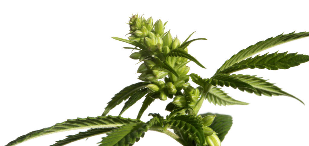
A cannabis plant against the white background

It is thought that dioecy confers a selective advantage in certain plant populations as it maximises the chances of genetic recombination. In hermaphrodite or monoecious plants, both male and female sexual organs are produced by the same plant. If it self-pollinates it will produce offspring whose DNA is identical to the parent. This lack of variation can very quickly lead to inbreeding and weakened genetic health in a population.

However, many hermaphroditic species have built-in genetic mechanisms that preclude self-pollination, a condition known as self-incompatibility.

It appears that monoecy and dioecy evolve in plants if the genetic mechanism for self-incompatibility has been lost (although they are each only present in around 7% of species, the capability has evolved independently in around 38% of all genera). There are several examples of dioecious plants expressing monoecious phenotypes in response to environmental pressures. But where those populations have again spread to more favourable locations, they gradually tend more towards dioecy.

This is an effective mechanism to ensure cross-pollination and genetic diversity in the absence of self-incompatibility.

This is borne out by cannabis, which has several monoecious strains and a great tendency to produce monoecious plants in dioecious populations, particularly in times of stress, and is entirely capable of self-pollination. In cannabis, numbers of males can drastically diminish for short periods in adverse conditions, but a strong and healthy male population is the default method to ensure long-term health and viability of the species.
 

acespicoli

Well-known member
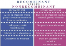
Recombinant and non-recombinant can refer to DNA, proteins, cells, or organisms that are either made by combining genetic material from different sources or are similar to the original or parental DNA:


  • Recombinant
    DNA, proteins, cells, or organisms that are made by combining genetic material from different sources. Recombinant DNA is created by connecting at least two distinct strands of DNA, and the plasmid vector is inserted with foreign DNA. Recombinant substances are made in laboratories and are being studied for many uses, including cancer treatment.


  • Non-recombinant
    DNA, proteins, cells, or organisms that are similar to the original or parental DNA. Non-recombinant DNA does not exhibit any genetic recombination, and there is no insertion of foreign DNA. Non-recombinant DNA exhibits little genetic variation and so has no impact on evolution.
The blue-white screening system is used to distinguish nonrecombinant DNA molecules from recombinants.

The blue-white screening system is a molecular biology technique that identifies recombinant bacteria by distinguishing between bacterial colonies that contain a cloning vector with a DNA insert and those that contain an empty vector:


  • How it works
    The system uses the activity of the enzyme β-galactosidase, which cleaves lactose into glucose and galactose. Plasmid vectors used in cloning carry a fragment of the lacZ gene. When plated on media containing X-Gal, the β-gal enzyme cleaves the colorless X-Gal and forms a bright blue precipitate, turning the bacterial colony blue.


  • What it indicates
    Blue colonies on a culture plate indicate that the bacterial cells have taken up a vector with an intact lacZα gene, meaning there is no foreign DNA inserted into the gene.


  • Why it's useful
    Blue-white screening is a convenient and powerful way to distinguish between colonies with and without a DNA insert. It's a widely used technique that can help make results from restriction enzyme digestion and ligation of DNA into a plasmid vector easier to discern.
 

Attachments

  • 1730216251473.png
    1730216251473.png
    235.7 KB · Views: 16

acespicoli

Well-known member
1730216531510.png

a In this schematic example, the MAGIC founders are genotyped more densely and confidently than the MAGIC recombinant inbred lines. The observed genotypes in the recombinant inbred lines (white) can be used to infer the ancestry mosaics (background colours) from which unobserved genotypes (black) can be imputed. Two examples of ancestry mosaics reconstructed from low-coverage sequence data in b Arabidopsis thaliana and c bread wheat. In b, the ancestry mosaic is estimated using the Reconstruction program (http://mtweb.cs.ucl.ac.uk/mus/www/19genomes/MAGICseq.htm), and accuracy is assessed as the fraction of mismatches in each block between the inferred founder haplotypes and calls derived directly from low-coverage sequencing data. In c, the inferred ancestry proportion probabilities (dosages) are emitted after imputation using the software, STITCH (Davies et al. 2016).

Scott, M.F., Ladejobi, O., Amer, S. et al. Multi-parent populations in crops: a toolbox integrating genomics and genetic mapping with breeding. Heredity 125, 396–416 (2020). https://doi.org/10.1038/s41437-020-0336-6

Download citation

  • Received27 January 2020
  • Revised16 June 2020
  • Accepted16 June 2020
  • Published03 July 2020
  • Issue DateDecember 2020
  • DOIhttps://doi.org/10.1038/s41437-020-0336-6
1730216741337.png
 

acespicoli

Well-known member
Double-stranded DNA (dsDNA) is the primary form of genetic material in most organisms and is made up of two polynucleotide chains that wind around each other to form a helix:


Double-stranded DNA
StructureTwo strands that run in opposite directions and are held together by hydrogen bonds between the bases
BasesAdenine (A) pairs with thymine (T), and cytosine (C) pairs with guanine (G)
BackbonesAlternating sugar (deoxyribose) and phosphate groups
ReplicationProkaryotic chromosomes have a single replication origin, while eukaryotic chromosomes have multiple replication origins
The double-stranded structure of DNA helps maintain genome stability by providing a template for repairing damage and replication mistakes. However, DNA double-strand breaks (DSBs) can occur in cells, which can lead to cell death, mutations, or cellular senescence.

1730217077420.png

Microspores are land plant spores that develop into male gametophytes, whereas megaspores develop into female gametophytes.[1] The male gametophyte gives rise to sperm cells, which are used for fertilization of an egg cell to form a zygote. Megaspores are structures that are part of the alternation of generations in many seedless vascular cryptogams, all gymnosperms and all angiosperms. Plants with heterosporous life cycles using microspores and megaspores arose independently in several plant groups during the Devonian period.[2] Microspores are haploid, and are produced from diploid microsporocytes by meiosis.[3]

Zygote

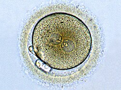
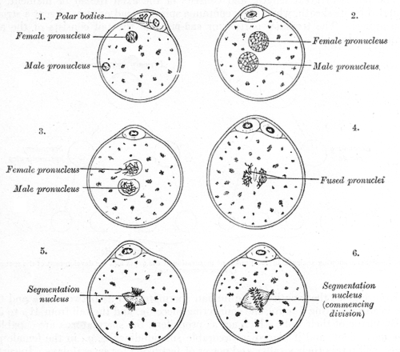
Zygote formation: egg cell after fertilization with a sperm.
The male and female pronuclei are converging, but the genetic material is not yet united.


The process of fertilization in the ovum of a mouse
A pronucleus (pl.: pronuclei) denotes the nucleus found in either a sperm or egg cell during the process of fertilization. The sperm cell undergoes a transformation into a pronucleus after entering the egg cell but prior to the fusion of the genetic material of both the sperm and egg. In contrast, the egg cell possesses a pronucleus once it becomes haploid, not upon the arrival of the sperm cell. Haploid cells, such as sperm and egg cells in humans, carry half the number of chromosomes present in somatic cells, with 23 chromosomes compared to the 46 found in somatic cells. It is noteworthy that the male and female pronuclei do not physically merge, although their genetic material does. Instead, their membranes dissolve, eliminating any barriers between the male and female chromosomes, facilitating the combination of their chromosomes into a single diploid nucleus in the resulting embryo, which contains a complete set of 46 chromosomes.


Plant breeding relies on crossing-over to create novel combinations of alleles needed to confer increased productivity and other desired traits in new varieties. However, crossover (CO) events are rare, as usually only one or two of them occur per chromosome in each generation. In addition, COs are not distributed evenly along chromosomes. In plants with large genomes, which includes most crops, COs are predominantly formed close to chromosome ends, and there are few COs in the large chromosome swaths around centromeres. This situation has created interest in engineering CO landscape to improve breeding efficiency. Methods have been developed to boost COs globally by altering expression of anti-recombination genes and increase CO rates in certain chromosome parts by changing DNA methylation patterns. In addition, progress is being made to devise methods to target COs to specific chromosome sites. We review these approaches and examine using simulations whether they indeed have the capacity to improve efficiency of breeding programs. We found that the current methods to alter CO landscape can produce enough benefits for breeding programs to be attractive. They can increase genetic gain in recurrent selection and significantly decrease linkage drag around donor loci in schemes to introgress a trait from unimproved germplasm to an elite line. Methods to target COs to specific genome sites were also found to provide advantage when introgressing a chromosome segment harboring a desirable quantitative trait loci. We recommend avenues for future research to facilitate implementation of these methods in breeding programs.

Proc Natl Acad Sci U S A
. 2023 Mar 27;120(14):e2205785119. doi: 10.1073/pnas.2205785119

Exploring impact of recombination landscapes on breeding outcomes​


Genome Res
. 2019 Jan;29(1):146–156. doi: 10.1101/gr.242594.118

A physical and genetic map of Cannabis sativa identifies extensive rearrangements at the THC/CBD acid synthase loci​


THCAS and CBDAS (which determine the drug vs. hemp chemotype) are contained within large (>250 kb) retrotransposon-rich regions that are highly nonhomologous between drug- and hemp-type alleles and are furthermore embedded within ∼40 Mb of minimally recombining repetitive DNA. The chromosome structures are similar to those in grains such as wheat, with recombination focused in gene-rich, repeat-depleted regions near chromosome ends. The physical and genetic map should facilitate further dissection of genetic and molecular mechanisms in this commercially and medically important plant.
>

1730217959622.png

Comparison of physical and genetic distance in Cannabis sativa and arrangement of sequence features on chromosomes. (A) Median values are indicated for all metacentric linkage groups (Chromosomes 5, 9, and 10 are excluded), scaled to the same physical length. Black points indicate the median increase in genetic distance every 1/100th of the physical distance. Shaded histograms superimposed show density of repeat sequences. Density of genes and GC content are also indicated by blue and purple lines. (B) Values for Chromosome 6, which contains the THCAS/CBDAS loci; here, black points are the representative of individual scaffolds.

A combined genetic and physical map reveals that genes and recombination events are concentrated near chromosome ends​

>

Recombinant CBCAS enzyme expression and purification​

The culture with the highest CBCAS activity was selected for scaled up production. One milliliter of the initial culture was used to inoculate two 40 mL BMG cultures, which were grown for 2 d at 37°C. These cultures were then used to initiate two 400 mL modified BMM cultures that were buffered with 10 mM HEPES (pH 7) and were supplemented with riboflavin at 20 mg/L. These cultures were grown at 20°C with shaking at 100 RPM for 5 d, with methanol added to 1% by volume each day. The cultures were then clarified by centrifugation, and the resulting media were filtered and passed over two Bio-scale Mini CHT hydroxyapatite cartridges (Bio-Rad) at a flow rate of 1.5 mL/min at 4°C. The cartridges were then attached in series to an AKTA FPLC system (GE Healthcare) and eluted with a 75-mL linear gradient from 5 mM sodium phosphate (pH 7) to 500 mM sodium phosphate (pH 7). Active fractions were pooled, concentrated with a 30 kDa cutoff Centricon filter (Millipore), and buffer exchanged into 20 mM citrate (pH 4.7) using a PD10 column (GE Healthcare). The resulting fraction was then injected onto a MonoS 5/50 cation exchange column (GE Healthcare) and eluted with a 40-mL linear gradient of 20 mM citrate (pH 4.7) to 20 mM citrate (pH 4.7) + 500 mM NaCl. Active fractions were pooled, concentrated with a 30 kDa cutoff Centricon filter, and injected onto a Hiload 26/60 Superdex 200 size exclusion column (GE Healthcare). Proteins were eluted with a single column volume of 20 mM citrate (pH 5.0) + 150 mM NaCl. Throughout the purification, 1/10th volume of each fraction was retained for analysis to judge purity. Protein was isolated from each fraction using 15 µL of StrataClean resin (Stratagene) and analyzed by SDS PAGE.

Methods​

Plant cultivation and gDNA isolation​

A female PK plant, produced through multiple vegetative propagation generations from the original source plant used to produce the draft C. sativa genome (van Bakel et al. 2011), was pollinated by a male FN plant in an indoor growth chamber. Seeds produced from this cross were germinated under standard conditions and grown to seedling stage. gDNA was isolated from young leaves using a GenElute Genomic Miniprep Kit (Sigma-Aldrich). The secure facilities used for plant growing were licensed by Health Canada.
1730218402659.png

Sequence heterogeneity of cannabidiolic- and tetrahydrocannabinolic acid-synthase in Cannabis sativa L. and its relationship with chemical phenotype​

Author links open overlay panelChiara Onofri a, Etienne P.M. de Meijer b, Giuseppe Mandolino a

https://doi.org/10.1016/j.phytochem.2015.03.006Get rights and content
 

Attachments

  • 1730218259365.png
    1730218259365.png
    1 MB · Views: 20
  • 1730218303700.png
    1730218303700.png
    571.2 KB · Views: 16
Last edited:

acespicoli

Well-known member

Complex Patterns of Cannabinoid Alkyl Side-Chain ...

1730218571124.png
National Institutes of Health (NIH) (.gov)
https://www.ncbi.nlm.nih.gov › articles › PMC6684623


by MT Welling · 2019 · Cited by 19 — While the presence of tandem THCAS as well as CBDAS arrays would imply oligogenic inheritance, genepool representative germplasm segregate ...

SNP in Potentially Defunct Tetrahydrocannabinolic Acid ...

1730218571157.png
National Institutes of Health (NIH) (.gov)
https://www.ncbi.nlm.nih.gov › articles › PMC7916091


by AR Garfinkel · 2021 · Cited by 28 — CBGA dominance is inherited as a single recessive gene, potentially governed by a non-functioning allelic variant of the THCA synthase.

Oligogenic inheritance is a pattern of inheritance where a trait is influenced by a small number of genes, usually three or more:


  • Explanation
    Oligogenic inheritance is a middle ground between monogenic inheritance, where a single gene determines a trait, and polygenic inheritance, where many genes and environmental factors influence a trait.
  • Characteristics
    Oligogenic conditions often have a wide range of phenotypic spectrum and variable severity. Mutations in multiple genes can interact with each other and environmental factors to influence disease progression and phenotypic variability.


    • Examples
      Some conditions associated with oligogenic inheritance include:
        • Breast cancer

        • Hirschsprung disease

        • Congenital hypogonadotropic hypogonadism

        • Familial hypercholesterolaemia

        • Severe adult obesity
    • Testing
      Oligogenic inheritance tests can detect risk genes and their interactions in developmental comorbidities and congenital heart defects.
    • 1730218716364.png

      De Luca, C., Buratti, A., Krauke, Y. et al. Investigating the effect of polarity of stationary and mobile phases on retention of cannabinoids in normal phase liquid chromatography.
    • Anal Bioanal Chem 414, 5385–5395 (2022). https://doi.org/10.1007/s00216-021-03862-y
      • DOIhttps://doi.org/10.1007/s00216-021-03862-y
 
Last edited:

acespicoli

Well-known member
1730221228345.png

In DNA, hydrogen bonds are the intermolecular forces that hold together the two strands of the double helix by connecting complementary nitrogenous bases (like adenine to thymine and cytosine to guanine) on opposite strands, essentially acting as the "rungs" of the DNA ladder; these bonds are crucial for maintaining the stability of the DNA molecule.


Key points about hydrogen bonds in DNA:
  • Base pairing:
    Adenine always pairs with thymine forming two hydrogen bonds, while cytosine pairs with guanine forming three hydrogen bonds.
    • Stability:
      These hydrogen bonds between base pairs provide the necessary stability for the DNA double helix structure.

    • Location:
      The hydrogen bonds occur between the nitrogenous bases on the interior of the DNA molecule.
1730221426950.png
 
Last edited:

acespicoli

Well-known member

DNA is made up of nucleotides​

DNA (deoxyribonucleic acid) is a nucleic acid biomolecule made up of subunits called nucleotides. Nucleotides make up an organism’s genetic information—certain stretches of nucleotides in a DNA molecule are genes, which encode the proteins that affect an organism’s traits. In addition, some stretches of nucleotides are involved in regulating when and how strongly those genes are expressed (used to make proteins).
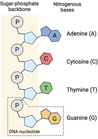
A DNA nucleotide has three components: a 5-carbon sugar (deoxyribose), a phosphate group, and a nitrogenous base. There are four types of nitrogenous bases found in DNA nucleotides: adenine, guanine, cytosine, or thymine. Scientists refer to nucleotides by the first letter of their base (A, G, C, and T, respectively).
Nucleotides are joined together by
covalent bonds
, which form between the deoxyribose sugar of one nucleotide and the phosphate group of the next. This arrangement makes an alternating chain of sugars and phosphate groups—a structure known as the sugar-phosphate backbone. Nucleotides can join together in any order, which means that any sequence of bases is possible.

Four bonded D N A nucleotides are shown. Each one consists of a phosphate group designated by a P, and 5-carbon sugar, and a nitrogenous base. Each nucleotide has a differently labeled nitrogenous base: A (adenine), C (cytosine), T (thymine), and G (guanine).

The structure of a DNA strand. DNA is made up of four types of nucleotides: adenine (A), cytosine (C), thymine (T), and guanine (G). Each nucleotide consists of a phosphate group (P), a deoxyribose sugar, and a nitrogenous base. The phosphate of one nucleotide is covalently bonded to the sugar of the next, forming a sugar-phosphate backbone. Created with Biorender.com.

DNA forms a double helix​

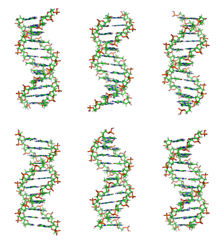
In the cell, DNA takes on the form of a double helix, which consists of two DNA strands that wind around each other like a twisted ladder. The sugar phosphate backbones of the DNA strands are on the outside of the double helix, forming the sides of the ladder. The nitrogenous bases point inward towards one another, forming the rungs of the ladder.

A video shows a rotating DNA double helix.

A video shows the three-dimensional shape of a DNA double helix. The double helix consists of two DNA strands wound around each other, with their sugar-phosphate backbones on the outside of the helix, and their nitrogenous bases pointing inward. Image credit: "DNA orbit animated" by Richard Wheeler (Zephyris), CC BY-SA 3.0
A DNA double helix has three primary features that are important for its function: antiparallel strands, hydrogen bonds between strands, and complementary base pairing.

Antiparallel strands​

Each strand of DNA in the double helix has directionality—that is, it has two ends that are different from each other. At one end, we find the phosphate group of the first nucleotide in the chain. This is called the 5’ end. At the other end, we find the deoxyribose sugar of the last nucleotide in the chain. This is called the 3’ end.
When the two strands of DNA come together in a double helix, the strands are antiparallel. This means that they point in opposite directions—the 5' end of one strand aligns with the 3' end of its partner strand, and vice versa.

Hydrogen bonds between strands​

The two strands of DNA in a double helix are joined by
hydrogen bonds
, which form between the nitrogenous bases of nucleotides on opposite strands. Each individual hydrogen bond is relatively weak compared to the bonds that hold together the sugar-phosphate backbone. However, collectively the bonds provide enough force to hold the strands together while still allowing the strands to be separated during DNA replication (when DNA is copied).

Complementary base pairing​

The hydrogen bonds between DNA strands occur between specific pairs of nitrogenous bases: T only pairs with A, and C only pairs with G. This specificity comes from the shapes of the bases and their chemical properties. Because the DNA strands are matched according to these base pairing rules, the strands are said to be complementary.

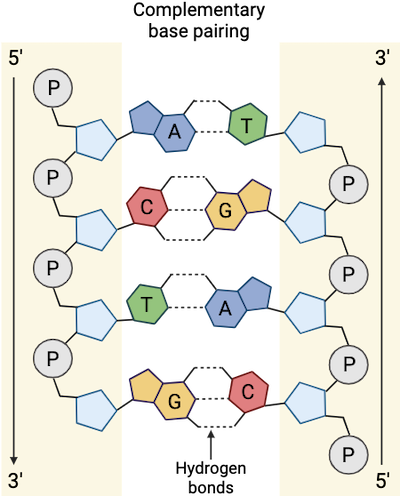
A model shows a flattened, double stranded D N A molecule with paired nitrogenous bases. A (adenine) on one strand is paired with T (thymine) on the other strand, and C (cytosine) on one strand is paired with G (guanine) on the other strand. The paired nitrogenous bases are joined by hydrogen bonds, which are represented by dotted lines. One of the strands is shown going in the 5 prime to 3 prime direction, and the other strand is antiparallel to it, going in the 3 prime to 5 prime direction.

In a DNA double helix, the DNA strands are antiparallel and attached via hydrogen bonding between nitrogenous bases. The double helix also shows complementary base pairing: A always pairs with T, and C always pairs with G. Created with Biorender.com.
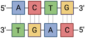
Complementary base pairing means that the two strands of a DNA double helix have a predictable relationship to each other. For instance, if we know that the sequence of one strand is 5’-ACTG-3’, then the complementary strand must have the sequence 3’-TGAC-5’. This allows each base to match up with its partner:

Two antiparallel D N A strands show complementary base pairing. A is always paired with T and C is always paired with G.

Created with Biorender.com.
When scientists write out the sequence of a gene, they typically write only the nucleotides from one strand, in the 5’ to 3’ direction. So, if the above sequence were part of a gene, it would be written as ACTG.
 

acespicoli

Well-known member
Rules of base pairing
  • A pairs with T (or U if RNA)
  • G pairs with C
Because of the structure of the bases, A can only form hydrogen bonds with T, and G can only form hydrogen bonds with C (remember Chargaff’s Rules). Each strand is therefore said to be complementary to the other, and so each strand also contains enough information to act as a template for the synthesis of the other. This complementary redundancy is important in DNA replication and repair. If the sequence of one strand is AATTGGCC, the complementary strand would have the sequence TTAACCGG. During DNA replication, each strand is copied, resulting in a daughter DNA double helix containing one parental DNA strand and a newly synthesized strand.

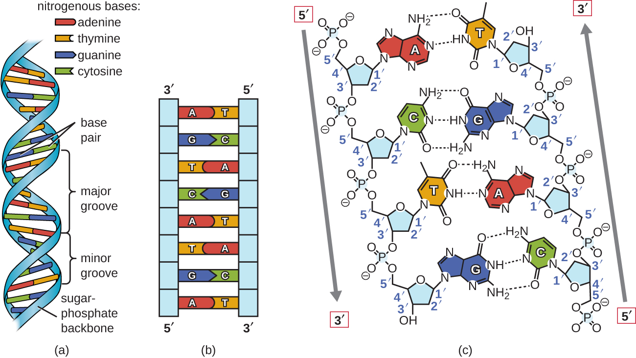
a) A diagram of DNA shown as a double helix (a twisted ladder). The outside of the ladder is a blue ribbon labeled “sugar phosphate backbone”. The rungs of the ladder are labeled “base pair” and are either red and yellow or green and blue. Red indicates the nitrogenous base adenine. Yellow indicates the nitrogenous base thymine. Blue indicates the nitrogenous base guanine. Green indicates the nitrogenous base cytosine. The ladder twists so that there are wide spaces (called major grooves) and narrow spaces (called minor grooves) between the twists. B) A different diagram of DNA showing it as a straight ladder. This makes it easier to see the bases (which can now be labeled with the letters A, T, C or G directly on the image. The left strand has a 3-prime at the top and a 5-prime at the bottom. The right strand has a 5-prime at the top and a 3-prime at the bottom. C) Another diagram of DNA showing a much shorter segment which allows the chemical structures to be seen more clearly. The strands show that the phosphate group is always between carbon 3 of one nucleotide and carbon 5 of the next. The two strands are connected with dotted lines indicating hydrogen bonds. The A-T bond has 2 hydrogen bonds and C-G has 3 hydrogen bonds. The negative charge of the phosphates is also apparent.
Figure 1.1.21.1.2:Watson and Crick proposed the double helix model for DNA. (a) The sugar-phosphate backbones are on the outside of the double helix and purines and pyrimidines form the “rungs” of the DNA helix ladder. (b) The two DNA strands are antiparallel to each other. (c) The direction of each strand is identified by numbering the carbons (1 through 5) in each sugar molecule. The 5ʹ end is the one where carbon #5 is not bound to another nucleotide; the 3ʹ end is the one where carbon #3 is not bound to another nucleotide and has a free hydroxyl group. (OpenStax-CNX)


3D structure of a DNA double helix
Spin the double helix to see the orientation of the sugars and phosphates in the backbone (ribbon in the model), the base pairs, major and minor grooves! (PDB ID = 1bna https://www.rcsb.org/3d-view/1BNA)
GLmol

Implications of DNA structure​

As for most biological molecules, the structure is important to the function, and the function of DNA is to contain information. Important properties that are derived from the DNA structure are:
  • A complementary strand can always be synthesized from a single strand, due to the arrangement of hydrogen bonds between GC and AT bases.
  • Hydrogen bonds stabilize the double helix, but can be broken when DNA needs to be accessed.
  • The order of bases contains the information needed to code for amino acids in proteins during translation.
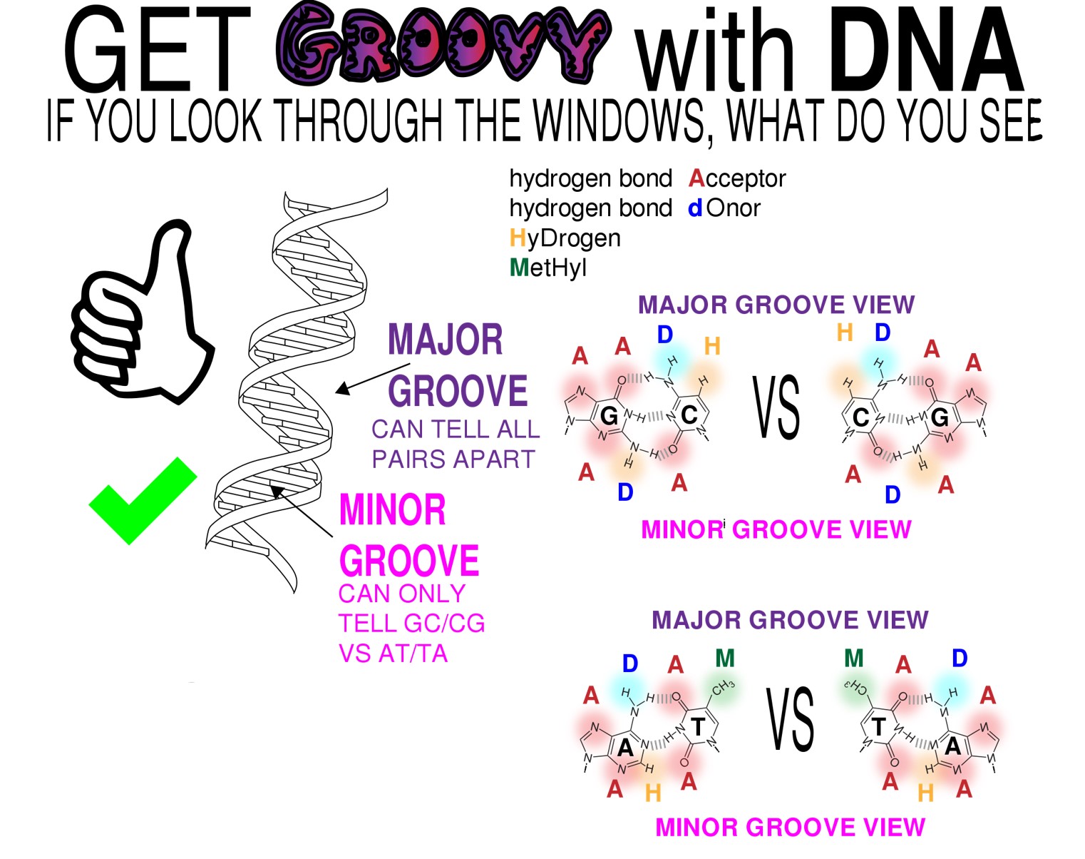
  • Even sequences of DNA that do not encode amino acids can still provide information by interacting with proteins that function in DNA packaging and regulation. The major and minor grooves of DNA may determine which sequences are visible to DNA interacting proteins.
DNA groovy biochem life cropped.jpg


Figure 1.1.31.1.3: The significance of major and minor grooves in a DNA double helix. A DNA double helix twists in a right-handed fashion, just as the fingers on the right hand are "pointing" to the right when the right hand forms a "thumbs up." The predominant structure of a double helix results in major and minor grooves. The bases within the double helix interact with each other via hydrogen bonds, but the different bases pairs have different combinations atoms exposed in the major grooves. Proteins that recognize DNA sequences often do so by interacting with particular combinations of base pairs in major grooves based on these exposed atoms. In the minor grooves AT and TA base pairs appear the same , likewise GC and CG look the same in the minor groove. (Copyright By Biochemlife - Own work, CC BY-SA 4.0, https://commons.wikimedia.org/w/inde...curid=73107644)


1730222279625.png
 

acespicoli

Well-known member
The Yang cycle is a series of reactions that recycles 5′-methylthioadenosine (MTA) into methionine in plants, bacteria, and yeast. It's a vital part of many metabolic pathways that support plant growth and development.


Here are some details about the Yang cycle:
 

acespicoli

Well-known member

Biological function and applications.​

doi: https://doi.org/10.1101/2023.04.28.538750
💥 ACC Synthase is the key, rate limiting step in ethylene synthesis. Because the up-regulation of ACC-Synthase is what induces fruit ripening and often spoilage there is more research being done on the regulatory mechanisms and biosynthetic pathways of ethylene to avoid spoilage.
1730255182656.png

1730255223994.png
1730255234947.png

1730255476770.png


Last edited:

acespicoli

Well-known member
1730255614430.png

1730255809857.png
Figure 2 Ethylene signaling transduction pathway. Model of the ethylene signal transduction. Under the mediation of RAN1, which transports copper to ethylene receptors, the presence of ethylene causes the loss of phosphorylation (P) of ethylene receptors (ETRs) at the membrane level (ER) (Binder et al., 2010); following this, the receptor–CTR1 complex is inactivated, the delivery of phosphate groups from CTR1 to EIN2 becomes incapable, and then EIN2 is cleaved and partly enters the nucleus to activate EIN3/EIL1 (Wen et al., 2012). Subsequently, EIN3/EIL1 binds to a conserved motif known as the EIN3 binding site (EBS), which is present within the promoters of ERF1, and this ultimately activates ERF1, which binds to the GCC box in the promoters of many ethylene-inducible, ripening-related genes (Fujimoto et al., 2000). Protein degradation of EIN3/EIL1 is regulated by EBF1/2 via the ubiquitin/26S proteasome pathway, and EIN5/XRN4 5′–3′ exoribonuclease mediated control of EBF1/2 mRNA levels (Potuschak et al., 2003; Olmedo et al., 2006).
1730255924236.png

Figure 3 Comparative ethylene evolution in representative climacteric and non-climacteric fruit models. (A) Ethylene evolution in the development of the tomato from the immature green to the red ripe stage (Zhang et al., 2009). IG, immature green (20 days after anthesis); MG, mature green (40 days after anthesis); B1, breaker (44 days after anthesis); B2, breaker (45 days after anthesis); T, turning (47 days after anthesis); P, pink (50 days after anthesis); MR, mature red (53 days after anthesis). (B) Ethylene evolution in the development of ‘Camarosa’ strawberry fruit during seven stages (Sun et al., 2013), SG (small green), BG (big green), DG (degreening), Wt (white), IR (initially red), PR (partially red), and FR (fully red), which occurred for 7, 15, 20, 23, 27, 31, and 35 days, respectively, after anthesis. (C) Ethylene evolution in the development of ‘Moldova’ grape fruit during contentious growth points of berry ripening (Xu et al., 2018). (D) Ethylene evolution in the development of attached ‘Valencia’ orange fruit during two growth stages (Katz et al., 2004). Stage I, the cell division stage, starts immediately after fruit set and lasts for approximately 90 days after full bloom (DAFB). Stage II, the cell expansion stage, during which fruit growth continues, mostly by cell expansion, extends until 150–180 DAFB.


Climacteric is a term that can refer to the transition from reproductive to non-reproductive
 

acespicoli

Well-known member
1730256158179.png

Figure 4 Phytohormone crosstalk between ethylene and other hormones in grape, strawberry, and citrus fruits. Note: The hormones and their related components involved in fruit ripening shown in the figure are abscisic acid (ABA), auxin (IAA),1-naphthaleneacetic acid (NAA), ethylene (C2H4), brassinosteroids (BRs), gibberellins (GAs), melatonin (MT) and jasmonates (JAs), tryptophan aminotransferase (TAR), auxin response factor (ARF), an auxin/indole-3-acetic acid (Aux/IAA) protein (IAA9), 9-cis-epoxycarotenoid dioxygenase 1 (NCED1), 1-aminocyclopropane-1-carboxylic acid synthase (ACS), and 1-aminocyclopropane-1-carboxylic acid oxidase (ACO). JA, ABA, BRs, and MT in the red fond have a positive effect on fruit ripening (Davies et al., 2006; Sun et al., 2010; Liu et al., 2011; Concha et al., 2013; Delgado et al., 2018; Garrido-Bigotes et al., 2018; Mansouri et al., 2021; Xia et al., 2021); Gas and IAA/NAA in the green fond had a negative effect on fruit ripening (Böttcher et al., 2011; Liu et al., 2011; Ma et al., 2021a, b; Tyagi et al., 2022); and jasmonate-activated fruit ripening is possibly associated with the stimulation of ethylene biosynthesis by an increase in ACO and ACS activities (Mukkun and Singh, 2009), while ABA, BRs, and MT can also stimulate ethylene production (Jiang and Joyce, 2003; Xu et al., 2018). C2H4 and IAA/NAA have a mutually reinforcing relationship. NAA can strongly upregulate ACS6 and ACO2 to improve ethylene biosynthesis (Ziliotto et al., 2012). In turn, the elevated concentrations of ethylene may lead to the induction of TAR expression, thus increasing the production of IAA (Böttcher et al., 2013). Ethylene also induces the transcription of VvNCED1 and the synthesis of ABA (Sun et al., 2010). The crosstalk between JAs and ABA, as well as GAs and IAA/NAA, also exists in non-climacteric fruit (Jung et al., 2014; Wang et al., 2015).
 

acespicoli

Well-known member
https://en.wikipedia.org/wiki/VSEPR_theory
1730256960898.png

Main-group elements​

For main-group elements, there are stereochemically active lone pairs E whose number can vary between 0 to 3. Note that the geometries are named according to the atomic positions only and not the electron arrangement. For example, the description of AX2E1 as a bent molecule means that the three atoms AX2 are not in one straight line, although the lone pair helps to determine the geometry.

ML7Pentagonal bipyramidal[10] ZrF3−
7Capped octahedral MoF−
7Capped trigonal prismatic TaF2−
7ML8Square antiprismatic[10] ReF−
8Dodecahedral Mo(CN)4−
8Bicapped trigonal prismatic ZrF4−
8ML9Tricapped trigonal prismatic ReH2−
9[13]: 254 Capped square antiprismatic
 

acespicoli

Well-known member

Molecular geometry​


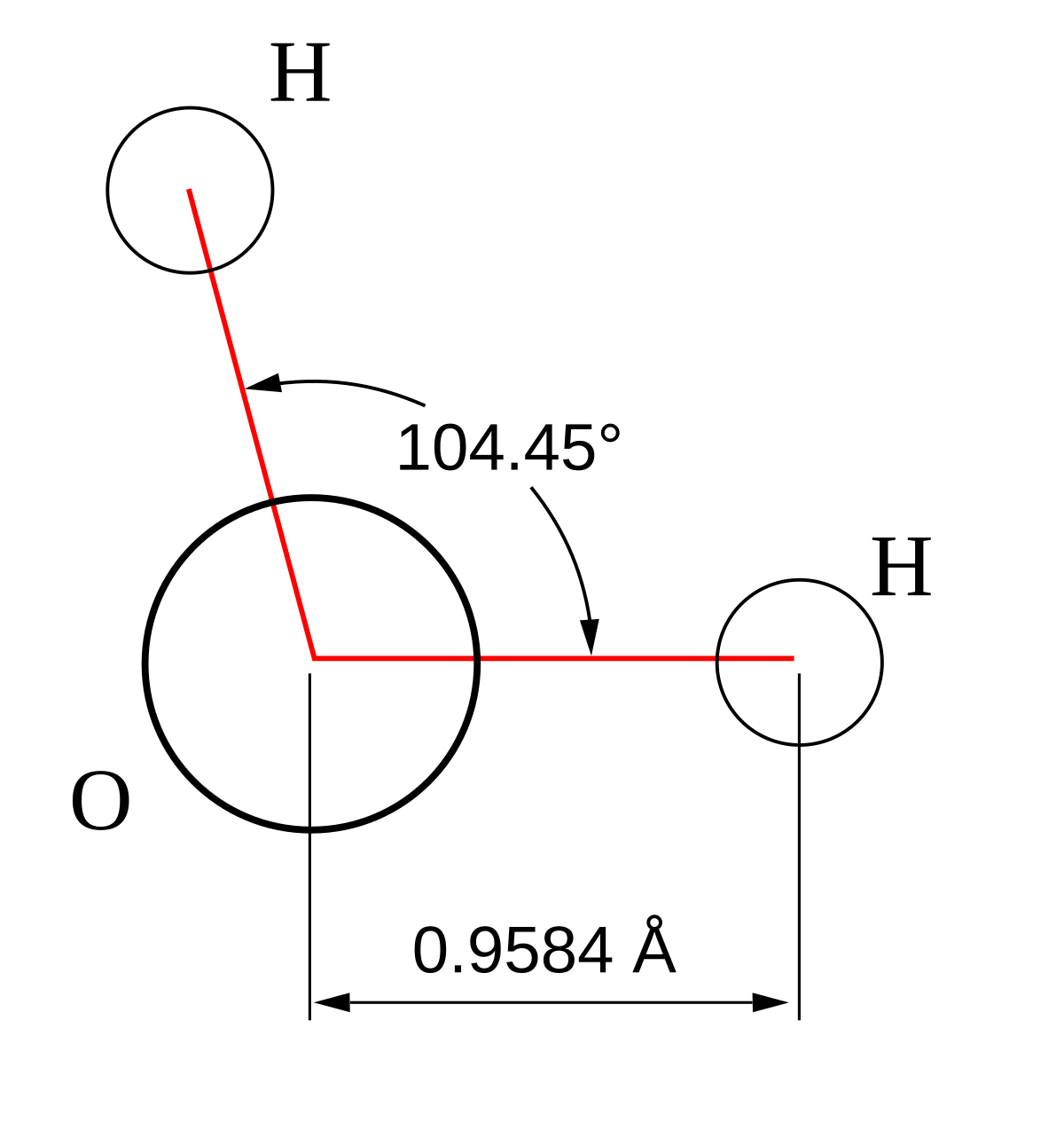
Geometry of the water molecule with values for O-H bond length and for H-O-H bond angle between two bonds
Molecular geometry is the three-dimensional arrangement of the atoms that constitute a molecule. It includes the general shape of the molecule as well as bond lengths, bond angles, torsional angles and any other geometrical parameters that determine the position of each atom.
Molecular geometry influences several properties of a substance including its reactivity, polarity, phase of matter, color, magnetism and biological activity.[1][2][3] The angles between bonds that an atom forms depend only weakly on the rest of molecule, i.e. they can be understood as approximately local and hence transferable properties.

Determination​

The molecular geometry can be determined by various spectroscopic methods and diffraction methods. IR, microwave and Raman spectroscopy can give information about the molecule geometry from the details of the vibrational and rotational absorbance detected by these techniques. X-ray crystallography, neutron diffraction and electron diffraction can give molecular structure for crystalline solids based on the distance between nuclei and concentration of electron density. Gas electron diffraction can be used for small molecules in the gas phase. NMR and FRET methods can be used to determine complementary information including relative distances,[4][5][6] dihedral angles,[7][8] angles, and connectivity. Molecular geometries are best determined at low temperature because at higher temperatures the molecular structure is averaged over more accessible geometries (see next section). Larger molecules often exist in multiple stable geometries (conformational isomerism) that are close in energy on the potential energy surface. Geometries can also be computed by ab initio quantum chemistry methods to high accuracy. The molecular geometry can be different as a solid, in solution, and as a gas.
The position of each atom is determined by the nature of the chemical bonds by which it is connected to its neighboring atoms. The molecular geometry can be described by the positions of these atoms in space, evoking bond lengths of two joined atoms, bond angles of three connected atoms, and torsion angles (dihedral angles) of three consecutive bonds.
 

acespicoli

Well-known member

Factors affecting ionic bond energy​

In ionic compounds, the electronegativity of the two atoms bonding together has a major effect on their bond energy.[14] The extent of this effect is described by the compound's lattice energy, where a more negative lattice energy corresponds to a stronger force of attraction between the ions. Generally, greater differences in electronegativity correspond to stronger ionic bonds. For example, the compound sodium chloride (NaCl) has a lattice energy of -786 kJ/mol with an electronegativity difference of 2.23 between sodium and chlorine. Meanwhile, the compound sodium iodide (NaI) has a lower lattice energy of -704 kJ/mol with a similarly lower electronegativity difference of 1.73 between sodium and iodine.[15][16]

See also​

Bond-dissociation energy
 

acespicoli

Well-known member
Front. Plant Sci., 05 June 2024
Sec. Plant Breeding
Volume 15 - 2024 | https://doi.org/10.3389/fpls.2024.1412079

Why not XY? Male monoecious sexual phenotypes challenge the female monoecious paradigm in Cannabis sativa L.


Current evidence suggests partial recombination is possible between the X and Y chromosomes in the male parent due to a pseudo autosomal region on the sex chromosomes (Peil et al., 2003).
 

acespicoli

Well-known member
Peil, A., Flachowsky, H., Schumann, E. et al. Sex-linked AFLP markers indicate a pseudoautosomal region in hemp (Cannabis sativa L.). Theor Appl Genet 107, 102–109 (2003). https://doi.org/10.1007/s00122-003-1212-5

Download citation

  • Received30 April 2002
  • Accepted18 November 2002
  • Issue DateJune 2003
  • DOIhttps://doi.org/10.1007/s00122-003-1212-5
1730389467609.png


While many people will sell feminized seed, some will even offer a clone.
1730390114950.png

Current evidence suggests partial recombination is possible between the X and Y chromosomes in the male parent due to a pseudo autosomal region on the sex chromosomes (Peil et al., 2003).

Inheritance and function​

Normal male therian mammals have two copies of these genes: one in the pseudoautosomal region of their Y chromosome, the other in the corresponding portion of their X chromosome. Normal females also possess two copies of pseudoautosomal genes, as each of their two X chromosomes contains a pseudoautosomal region. Crossing over between the X and Y chromosomes is normally restricted to the pseudoautosomal regions; thus, pseudoautosomal genes exhibit an autosomal, rather than sex-linked, pattern of inheritance. So, females can inherit an allele originally present on the Y chromosome of their father.

The function of these pseudoautosomal regions is that they allow the X and Y chromosomes to pair and properly segregate during meiosis in males.[9]

The linked frequency of crossing over between two gene loci (markers) is the crossing-over value. For fixed set of genetic and environmental conditions, recombination in a particular region of a linkage structure (chromosome) tends to be constant and the same is then true for the crossing-over value which is used in the production of genetic maps.[6][7]

Origin in cell biology​

Crossover implies the exchange of chromosomal segments between non-sister chromatids, in meiosis during the production of gametes. The effect is to assort the alleles on parental chromosomes, so that the gametes carry recombinations of genes different from either parent. This has the overall effect of increasing the variety of phenotypes present in a population.

The process of non-sister chromatid exchanges, including the crossover value, can be observed directly in stained cells, and indirectly by the presence or absence of genetic markers on the chromosomes. The visible crossovers are called chiasmata.

The large-scale effect of crossover is to spread genetic variations within a population, as well as genetic basis for the selection of the most adaptable phenotypes. The crossover value depends on the mutual distance of the genetic loci observed. The crossover value is equal to the recombination value or fraction when the distance between the markers in question is short.

1730390536961.png

An efficient RNA-seq-based​

segregation analysis identifies the sex chromosomes of Cannabis sativa​

DOI:10.1101/gr.251207.119
 
Last edited:

acespicoli

Well-known member
Backcrossing is a crossing of a hybrid with one of its parents or an individual genetically similar to its parent, to achieve offspring with a genetic identity closer to that of the parent. It is used in horticulture, animal breeding, and production of gene knockout organisms.

Backcrossed hybrids are sometimes described with acronym "BC"; for example, an F1 hybrid crossed with one of its parents (or a genetically similar individual) can be termed a BC1 hybrid, and a further cross of the BC1 hybrid to the same parent (or a genetically similar individual) produces a BC2 hybrid.[1]

Plants​

Advantages
If the recurrent parent is an elite genotype, at the end of the backcrossing programme, an elite genotype is recovered.
  • As no "new" recombination results, the elite combination is not lost.

Disadvantages​

It works poorly for quantitative traits.
  • It is more restricted for recessive traits.
  • In practice, sections of genome from the nonrecurrent parents are often still present and can have unwanted traits associated with them.
  • For very wide crosses, limited recombination may maintain thousands of "alien" genes within the elite cultivar.
  • Many backcrosses are required to produce a new cultivar, which can take many years.
 

acespicoli

Well-known member
the targeted removal or inactivation of a specific gene within an organism's genome.
This can be done through a variety of methods, including homologous recombination,

Mutations in genes that encode proteins involved in the processing of DNA often affect recombination frequency.


  • With very large pedigrees or with very dense genetic marker data, such as from whole-genome sequencing, it is possible to precisely locate recombinations. With this type of genetic analysis, a meiosis indicator is assigned to each position of the genome for each meiosis in a pedigree. The indicator indicates which copy of the parental chromosome contributes to the transmitted gamete at that position. For example, if the allele from the 'first' copy of the parental chromosome is transmitted, a '0' might be assigned to that meiosis. If the allele from the 'second' copy of the parental chromosome is transmitted, a '1' would be assigned to that meiosis. The two alleles in the parent came, one each, from two grandparents. These indicators are then used to determine identical-by-descent (IBD) states or inheritance states, which are in turn used to identify genes responsible for diseases.
    1730425168135.png


    Gene pyramiding is the simultaneous selection for and/or introduction of multiple genes during plant breeding.[1] Objectives of gene pyramiding includes 1) enhancing trait performance by combining two or more complementary genes, 2) remedying deficits by introgressing genes from other sources, 3) increasing the durability.[2] For example, pyramiding has been successfully demonstrated in Oryza sativa for rice blast, producing durable multi-race resistance simultaneously.[3] Pyramiding and Marker Assisted Selection can be combined as Marker-Assisted Pyramiding.[4] Gene stacking can be achieved a few different ways, and pyramiding is one of those methods.[5]


    For many years, gene mapping was limited to identifying organisms by traditional phenotypes markers. This included genes that encoded easily observable characteristics, such as blood types or seed shapes.


    Genetic markers are employed in genealogical DNA testing for genetic genealogy to determine genetic distance between individuals or populations. Uniparental markers (on mitochondrial or Y chromosomal DNA) are studied for assessing maternal or paternal lineages. Autosomal markers are used for all ancestry.


Latest posts

Latest posts

Top