What's new
  • ICMag and The Vault are running a NEW contest! You can check it here. Prizes are seeds & forum premium access. Come join in!

DIY Organic Potting Mix's for Grass - Ace Spicoli

acespicoli

Well-known member
In cellular biology, active transport is the movement of molecules or ions across a cell membrane from a region of lower concentration to a region of higher concentration—against the concentration gradient.

Biological perspective[edit]​

The first law gives rise to the following formula:[19]

flux=−�(�2−�1)
{\displaystyle {\text{flux}}={-P\left(c_{2}-c_{1}\right)}}

in which

  • P is the permeability, an experimentally determined membrane "conductance" for a given gas at a given temperature.
  • c2 − c1 is the difference in concentration of the gas across the membrane for the direction of flow (from c1 to c2).
Fick's first law is also important in radiation transfer equations. However, in this context, it becomes inaccurate when the diffusion constant is low and the radiation becomes limited by the speed of light rather than by the resistance of the material the radiation is flowing through. In this situation, one can use a flux limiter.

The exchange rate of a gas across a fluid membrane can be determined by using this law together with Graham's law.

Under the condition of a diluted solution when diffusion takes control, the membrane permeability mentioned in the above section can be theoretically calculated for the solute using the equation mentioned in the last section (use with particular care because the equation is derived for dense solutes, while biological molecules are not denser than water):[12]

{\displaystyle P=2A_{p}\eta _{tm}{\sqrt {D/(\pi t)}}}

where

  • {\displaystyle A_{P}}
    is the total area of the pores on the membrane (unit m2).
  • {\displaystyle \eta _{tm}}
    transmembrane efficiency (unitless), which can be calculated from the stochastic theory of chromatography.
  • D is the diffusion constant of the solute unit m2⋅s−1.
  • t is time unit s.
  • c2, c1 concentration should use unit mol m−3, so flux unit becomes mol s−1.
The flux is decay over the square root of time because a concentration gradient builds up near the membrane over time under ideal conditions. When there is flow and convection, the flux can be significantly different than the equation predicts and show an effective time t with a fixed value,[15] which makes the flux stable instead of decay over time. A critical time has been estimated under idealized flow conditions when there is no gradient formed.[15][17] This strategy is adopted in biology such as blood circulation.
 

acespicoli

Well-known member

Surface chemistry[edit]​


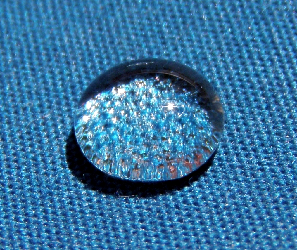
Contact angle of a liquid droplet wetted to a rigid solid surface.Young's equation: γLG ∙cos θ+ γSL= γSG.
One of the critical characteristics of a synthetic membrane is its chemistry. Synthetic membrane chemistry usually refers to the chemical nature and composition of the surface in contact with a separation process stream.[6] The chemical nature of a membrane's surface can be quite different from its bulk composition. This difference can result from material partitioning at some stage of the membrane's fabrication, or from an intended surface postformation modification. Membrane surface chemistry creates very important properties such as hydrophilicity or hydrophobicity (related to surface free energy), presence of ionic charge, membrane chemical or thermal resistance, binding affinity for particles in a solution, and biocompatibility (in case of bioseparations).[6] Hydrophilicity and hydrophobicity of membrane surfaces can be expressed in terms of water (liquid) contact angle θ. Hydrophilic membrane surfaces have a contact angle in the range of 0°<θ<90° (closer to 0°), where hydrophobic materials have θ in the range of 90°<θ<180°.


Wetting of a leaf.
The contact angle is determined by solving the Young's equation for the interfacial force balance. At equilibrium three interfacial tensions corresponding to solid/gas (γSG), solid/liquid (γSL), and liquid/gas (γLG) interfaces are counterbalanced.[6] The consequence of the contact angle's magnitudes is known as wetting phenomena, which is important to characterize the capillary (pore) intrusion behavior. Degree of membrane surface wetting is determined by the contact angle. The surface with smaller contact angle has better wetting properties (θ=0°-perfect wetting). In some cases low surface tension liquids such as alcohols or surfactant solutions are used to enhance wetting of non-wetting membrane surfaces.[6] The membrane surface free energy (and related hydrophilicity/hydrophobicity) influences membrane particle adsorption or fouling phenomena. In most membrane separation processes (especially bioseparations), higher surface hydrophilicity corresponds to the lower fouling.[6] Synthetic membrane fouling impairs membrane performance. As a consequence, a wide variety of membrane cleaning techniques have been developed. Sometimes fouling is irreversible, and the membrane needs to be replaced. Another feature of membrane surface chemistry is surface charge. The presence of the charge changes the properties of the membrane-liquid interface. The membrane surface may develop an electrokinetic potential and induce the formation of layers of solution particles which tend to neutralize the charge.


Explanation[edit]​

Figure 1: Contact angle for a liquid droplet on a solid surface
Adhesive forces between a liquid and solid cause a liquid drop to spread across the surface. Cohesive forces within the liquid cause the drop to ball up and avoid contact with the surface.

Solid–liquidLiquid–liquid
Fig. 2Contact angleDegree of
wetting
Interaction strength
Sθ = 0Perfect wettingStrongWeak
C0 < θ < 90°High wettabilityStrongStrong
WeakWeak
B90° ≤ θ < 180°Low wettabilityWeakStrong
Aθ = 180°Non-wettingWeakStrong
Figure 2: Wetting of different fluids: A shows a fluid with very little wetting, while C shows a fluid with more wetting. A has a large contact angle, and C has a small contact angle.
The contact angle (θ), as seen in Figure 1, is the angle at which the liquid–vapor interface meets the solid–liquid interface. The contact angle is determined by the balance between adhesive and cohesive forces. As the tendency of a drop to spread out over a flat, solid surface increases, the contact angle decreases. Thus, the contact angle provides an inverse measure of wettability.[7][8]

A contact angle less than 90° (low contact angle) usually indicates that wetting of the surface is very favorable, and the fluid will spread over a large area of the surface. Contact angles greater than 90° (high contact angle) generally mean that wetting of the surface is unfavorable, so the fluid will minimize contact with the surface and form a compact liquid droplet.

For water, a wettable surface may also be termed hydrophilic and a nonwettable surface hydrophobic. Superhydrophobic surfaces have contact angles greater than 150°, showing almost no contact between the liquid drop and the surface. This is sometimes referred to as the "Lotus effect". The table describes varying contact angles and their corresponding solid/liquid and liquid/liquid interactions.[9] For nonwater liquids, the term lyophilic is used for low contact angle conditions and lyophobic is used when higher contact angles result. Similarly, the terms omniphobic and omniphilic apply to both polar and apolar liquids.
 

acespicoli

Well-known member
does anyone have any references to research papers showing increasing terpenes with various organic materials.

as in a study done with terpenes tested first in a soilless media or hydro methods and then the same plant grown with various organic inputs.

fascinating subject and i will be following this thread for sure.

i have been adding small organic inputs to a soilless media in the PPK system and i am getting a stronger aroma profile. i would like to know whether i am actually increasing terpenes or simply injecting a substance that increases smell.

thus far i have not been able to find any research on the subject that actually quantifies this.
Chitin
View attachment 19033390
A close-up of the wing of a leafhopper; the wing is composed of chitin.


Front. Plant Sci., 19 September 2021
Sec. Plant Metabolism and Chemodiversity
Volume 12 - 2021 | https://doi.org/10.3389/fpls.2021.721986
"The role of cannabinoids in biotic stress tolerance is consistent with their elevated concentration in flowers where trichome densities are highest. In addition to reducing the risk of pest-related damage, cannabinoids also have antimicrobial properties."

Effects of chitin and chitosan on root growth, biochemical defense response and exudate proteome of Cannabis sativa​

10.1002/pei3.10106


Chicken feed oyster shell from tractor supply cheap...$
https://www.tractorsupply.com/tsc/product/manna-pro-oyster-shell-50-lb


Agricultural and horticultural use[edit]​

The agricultural and horticultural uses for chitosan, primarily for plant defense and yield increase, are based on how this glucosamine polymer influences the biochemistry and molecular biology of the plant cell. The cellular targets are the plasma membrane and nuclear chromatin. Subsequent changes occur in cell membranes, chromatin, DNA, calcium, MAP kinase, oxidative burst, reactive oxygen species, callose pathogenesis-related (PR) genes, and phytoalexins.[16]

Chitosan was first registered as an active ingredient (licensed for sale) in 1986.[17]

Natural biocontrol and elicitor[edit]​

In agriculture, chitosan is typically used as a natural seed treatment and plant growth enhancer, and as an ecologically friendly biopesticide substance that boosts the innate ability of plants to defend themselves against fungal infections.[18] The natural biocontrol active ingredients, chitin/chitosan, are found in the shells of crustaceans, such as lobsters, crabs, and shrimp, and many other organisms, including insects and fungi. It is one of the most abundant biodegradable materials in the world.[citation needed]

Degraded molecules of chitin/chitosan exist in soil and water. Chitosan applications for plants and crops are regulated in the USA by the EPA, and the USDA National Organic Program regulates its use on organic certified farms and crops.[19] EPA-approved, biodegradable chitosan products are allowed for use outdoors and indoors on plants and crops grown commercially and by consumers.[20]

In the European Union and United Kingdom, chitosan is registered as a "basic substance" for use as a biological fungicide and bactericide on a wide range of crops.[21][22]

The natural biocontrol ability of chitosan should not be confused with the effects of fertilizers or pesticides upon plants or the environment. Chitosan active biopesticides represent a new tier of cost-effective biological control of crops for agriculture and horticulture.[23] The biocontrol mode of action of chitosan elicits natural innate defense responses within plant to resist insects, pathogens, and soil-borne diseases when applied to foliage or the soil.[24] Chitosan increases photosynthesis, promotes and enhances plant growth, stimulates nutrient uptake, increases germination and sprouting, and boosts plant vigor. When used as a seed treatment or seed coating on cotton, corn, seed potatoes, soybeans, sugar beets, tomatoes, wheat, and many other seeds, it elicits an innate immunity response in developing roots which destroys parasitic cyst nematodes without harming beneficial nematodes and organisms.[25]

Agricultural applications of chitosan can reduce environmental stress due to drought and soil deficiencies, strengthen seed vitality, improve stand quality, increase yields, and reduce fruit decay of vegetables, fruits and citrus crops .[26] Horticultural application of chitosan increases blooms and extends the life of cut flowers and Christmas trees. The US Forest Service has conducted research on chitosan to control pathogens in pine trees[27][28] and increase resin pitch outflow which resists pine beetle infestation.[29]


NASA life support GAP technology with untreated beans (left tube) and ODC chitosan biocontrol-treated beans (right tube) returned from the Mir space station aboard the space shuttle – September 1997
Chitosan has a rich history of being researched for applications in agriculture and horticulture dating back to the 1980s.[30] By 1989, chitosan salt solutions were applied to crops for improved freeze protection or to crop seed for seed priming.[31] Shortly thereafter, chitosan salt received the first ever biopesticide label from the EPA, then followed by other intellectual property applications.

Chitosan has been used to protect plants in space, as well, exemplified by NASA's experiment to protect adzuki beans grown aboard the space shuttle and Mir space station in 1997 (see photo left).[32] NASA results revealed chitosan induces increased growth (biomass) and pathogen resistance due to elevated levels of β-(1→3)-glucanase enzymes within plant cells. NASA confirmed chitosan elicits the same effect in plants on earth.[33]

In 2008, the EPA approved natural broad-spectrum elicitor status for an ultralow molecular active ingredient of 0.25% chitosan.[34] A natural chitosan elicitor solution for agriculture and horticultural uses was granted an amended label for foliar and irrigation applications by the EPA in 2009.[26] Given its low potential for toxicity and abundance in the natural environment, chitosan does not harm people, pets, wildlife, or the environment when used according to label directions.[35][36][37] Chitosan blends do not work against bark beetles when put on a tree's leaves or in its soil.[38]


https://www.frontiersin.org/journals/marine-science/articles/10.3389/fmars.2018.00347/full
just dump a 50# bag in a a 6 cu sq ft run of my sig soil mix 🤷‍♂️
Chitin is broadly distributed in the shells of mollusks where it constitutes 3.5 wt%
 
Last edited:

acespicoli

Well-known member

The Rhizosphere - Roots, Soil and Everything In Between​

By: David H. McNear Jr. (Assistant Professor of Rhizosphere Science) © 2013 Nature Education


Citation: McNear Jr., D. H. (2013) The Rhizosphere - Roots, Soil and Everything In Between. Nature Education Knowledge 4(3):1


What is the Rhizosphere and how can understanding rhizosphere processes help feed the world and save the environment? This article will review the critical biogeochemical processes occurring at the plant root-soil interface.
Aa Aa Aa


McNear banner.


Meeting the Global Challenge of Sustainable Food, Fuel and Fiber Production​


Soil is one of the last great scientific frontiers (Science, 11 June, 2004) and the rhizosphere is the most active portion of that frontier in which biogeochemical processes influence a host of landscape and global scale processes. A better understanding of these processes is critical for maintaining the health of the planet and feeding the organisms that live on it (Morrissey et al., 2004). There is a small but concerted effort under way to harness the root system of plants in an attempt to increase yield potentials of staple food crops in order to meet the projected doubling in global food demand in the next 50 years (Zhang, et al. 2010; Gewvin, 2010). These efforts are being done in the face of a changing global climate and increasing global population which will inevitably require more productively grown food, feed and fiber on less optimal (and often infertile) lands; a condition already encountered in many developing countries (Tilman, et al, 2002). Meeting the global challenges of climate change and population growth with a better understanding and control of rhizosphere processes will be one of the most important science frontiers of the next decade for which a diverse, interdisciplinary trained workforce will be required.

The Rhizosphere Defined​

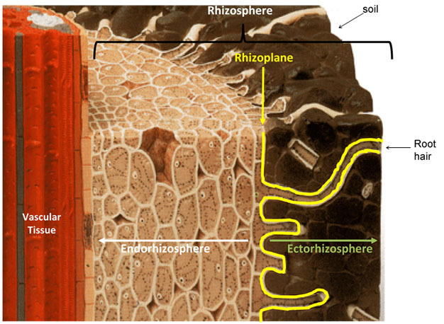
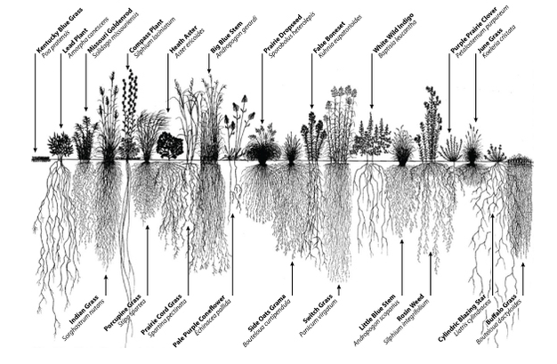
In 1904 the German agronomist and plant physiologist Lorenz Hiltner first coined the term "rhizosphere" to describe the plant-root interface, a word originating in part from the Greek word "rhiza", meaning root (Hiltner, 1904; Hartmann et al., 2008). Hiltner described the rhizosphere as the area around a plant root that is inhabited by a unique population of microorganisms influenced, he postulated, by the chemicals released from plant roots. In the years since, the rhizosphere definition has been refined to include three zones which are defined based on their relative proximity to, and thus influence from, the root (Figure 1). The endorhizosphere includes portions of the cortex and endodermis in which microbes and cations can occupy the "free space" between cells (apoplastic space). The rhizoplane is the medial zone directly adjacent to the root including the root epidermis and mucilage. The outermost zone is the ectorhizosphere which extends from the rhizoplane out into the bulk soil. As might be expected because of the inherent complexity and diversity of plant root systems (Figure 2), the rhizosphere is not a region of definable size or shape, but instead, consists of a gradient in chemical, biological and physical properties which change both radially and longitudinally along the root.
Schematic of a root section

Figure 1
Schematic of a root section showing the structure of the rhizosphere.
Modified from http://cse.naro.affrc.go.jp. View Terms of Use


Image showing the diversity of root system architecture in prairie plants

Figure 2
Image showing the diversity of root system architecture in prairie plants.
© 2012 Nature Education 1995 Conservation Research Institute, Heidi Natura. All rights reserved. View Terms of Use


Classification and Function of Root Derived Products​


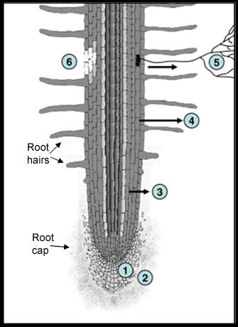
Newman (1985) examined a variety of plant species and estimated that roots can release anywhere from 10 to 250 mg C /g root produced or about 10-40% of their total photosynthetically fixed carbon. The C released is in both organic (e.g., low molecular weight organic acids) and inorganic (e.g., HCO3) forms, however, the organic forms are the most varied and can have the most influence on the chemical, physical and biological processes in the rhizosphere (Jones, et al., 2009). The composition and amount of the released compounds is influenced by many factors including plant type, climactic conditions, insect herbivory, nutrient deficiency or toxicity, and the chemical, physical and biological properties of the surrounding soil. The root products imparted to the surrounding soil are generally called rhizodeposits (Figure 3). Rhizodeposits have been classified based on their chemical composition, mode of release, or function but are classically defined (Rovaria, 1969) to include sloughed-off root cap and border cells, mucilage, and exudates.
Schematic of a root showing 6 major regions of rhizodeposits.

Figure 3
Schematic of a root showing the 6 major regions of rhizodeposits. 1 loss of cap and border cells, 2 loss of insoluble mucilage, 3 loss of soluble root exudates, 4 loss of volatile organic carbon, 5 loss of C to symbionts, 6 loss of C due to death and lysis of root epidermal and cortical cells.
© 2012 Nature Education Jones, D.L., Nguyen, C., and Finlay, R.D. Carbon flow in the rhizosphere: carbon trading at the soil-root interface. Plant Soil 321:5-33 (2009). All rights reserved. View Terms of Use


Roots exert a tremendous amount of pressure (>7kg/cm2 or ~100 psi) at the growing root tips in order to push their way through the soil. Helping to lubricate and protect the root during growth, root cap and epidermal cells secrete mucilage, a viscous, high molecular weight, insoluble, polysaccharide-rich material. Beyond lubrication, the mucilage also provides protection from desiccation, assists in nutrient acquisition, and most notably binds soil particles together forming aggregates which improve soil quality by increasing water infiltration and aeration. Also serving to protect the root tip, cells lining and capping the root meristem are programmed to release (slough-off) which helps to reduce frictional forces that would otherwise damage the root (Bengough and McKenzie, 1997). Interestingly, the cells that are sloughed off continue to function and secrete mucilage for several days and have been shown to attract beneficial microorganisms, serve as "bait" for root pathogens, and sequester toxic metals (e.g. Al3+) (Hawes et al., 2000).
Root exudates include both secretions (including mucilage) which are actively released from the root and diffusates which are passively released due to osmotic differences between soil solution and the cell, or lysates from autolysis of epidermal and cortical cells (Figure 4). The organic compounds released through these processes can be further divided into high and low molecular weight (HMW and LMW, respectively). By weight, the HMW compounds which are those complex molecules that are not easily used by microorganisms (e.g. mucilage, cellulose) make up the majority of C released from the root; however, the LMW compounds are more diverse and thus have a wider array of known or potential functions. The list of specific LMW compounds released from roots is very long, but can generally be categorized into organic acids, amino acids, proteins, sugar, phenolics and other secondary metabolites which are generally more easily used by microorganisms. The cocktail of chemicals released is influenced by plant species, edaphic and climactic conditions which together shape and are shaped by the microbial community within the rhizosphere. There is still very little known about the role that a majority of the LMW compounds play in influencing rhizosphere processes. A growing body of literature is beginning to lift the veil on the many functions of root exudates as a means of acquiring nutrients (e.g. acquisition of Fe and P), agents of invasiveness (i.e. allelopathy) or as chemical signals to attract symbiotic partners (chemotaxis) (e.g. rhizobia and legumes) or the promotion of beneficial microbial colonization on root surfaces (e.g. Bacillus subtilis, Pseudomonas florescence) (Bais, Park et al. 2004).
A branch root (red arrow) of buckwheat

Figure 4
A branch root (red arrow) of buckwheat (Fagopyron esculentum) frozen together with its surrounding soil and then imaged using scanning electron microscopy to reveal the intimate contact between the root and soil. Note the root hairs extending out and exploring the surrounding soil. Bar = 600 μm. b) Red arrows point to droplets of root exudates released from the tips of root hairs on the surface of broom corn (Sorghum sp.). Bar = 50 μm.
© 2012 Nature Education McGully, M. The rhizosphere: the key functional unit in plant/soil/microbial interactions in the field. implications for the understanding of allelopathic effects. Division of Plant Industry. Forth World Congress on Allelopathy. The Regional Institute Ltd. All rights reserved. View Terms of Use


Root Exudates and Mineral Nutrition​

Plants respond to nutrient deficiency by altering root morphology, recruiting the help of microorganisms and changing the chemical environment of the rhizosphere. Components in root exudates assist plants in accessing nutrients by acidifying or changing the redox conditions within the rhizosphere or directly chelating with the nutrient. Exudates can liberate nutrients via dissolution of insoluble mineral phases or desorption from clay minerals or organic matter where they are released into soil solution and can then be taken up by the plant. The nutrients most limiting to plant growth are nitrogen and phosphorus. Even thought 78% of the Earth's atmosphere is composed of nitrogen (N2 gas), it is in a form that is only utilizable by nitrogen-fixing organisms. As such, inorganic forms of N (NO3-,NH4+) that can be used by plants are added to soils. The availability of nitrogen in most soils is low because of the leaching losses of soluble nitrate (NO3-) with infiltrating rainwater, fixation of ammonium (NH4+) in clays and soil organic matter and bacterial denitrification. Plants respond differently depending on the form of nitrogen in the soil. Ammonium has a positive charge, and thus the plant expels one proton (H+) for every NH4+ taken up resulting in a reduction in rhizosphere pH. When supplied with NO3-, the opposite can occur where the plant releases bicarbonate (HCO3-) which increases rhizosphere pH. These changes in pH can influence the availability of other plant essential micronutrients (e.g., Zn, Ca, Mg).
Phosphate (PO43-), the form of P used by plants, is highly insoluble in soils, binding strongly to Ca, Al and Fe oxide, and soil organic matter rendering much of the P unavailable to plants. Under P deficiency, plants have evolved special mechanisms to obtain PO43- which depend on plant type (dicot vs. monocot), species and genotype. Plant roots can exude organic acids such as malic and citric acids into the rhizosphere which effectively reduced rhizosphere pH and solubilize P bound in soil minerals. Pigeon pea uses another mechanism by releasing piscidic acid in response to P deficiency which chelates Fe in FePO4, releasing the PO43- (Raghothama, 1999). Plants also liberate PO43- from organic sources by releasing enzymes such as acid phosphatase.
Iron deficiency elicits a response from plants which generally differs depending on whether the plant is a dicot or monocot. Dicots respond to Fe deficiency by releasing protons into the soil environment and increasing the reducing capacity of the rhizodermal cells. In monocots, Fe deficiency triggers the release of phytosiderophores such as mugienic acid which is a non-proteinogenic amino acid with extremely high affinity for Fe. The phytosiderophore chelates strongly with Fe and is then brought back to the root via diffusion where plasma membrane transporters specific to the chelated Fe shuttle it into the cells. Plants using the former method of Fe acquisition are called Strategy I and those using the latter Strategy II.

Key Microbe-Plant Relationships​

Rhizodeposits make the rhizosphere a desirable niche for microbial communities to proliferate. One teaspoon of bare or tilled soil contains more microorganisms than there are people on Earth, however, the rhizosphere can have 1000-2000 times that number (1010-1012 cells per gram rhizosphere soil) making it a pretty crowded place. Nutrient availability for microbial growth is higher in the rhizosphere compared to the bulk soil; however, many microbes are competing for these nutrients, some more successfully than others. The plant rhizosphere -microbe relationships that have received the most attention include those of Rhizobia bacteria and their symbiotic plant partners, mychorrhizal fungi associations, and beneficial plant growth promoting rhizobacteria (PGPR) each of which will be discussed in more detail below.


Legume-Rhizobia Symbiosis​


The observation that including leguminous plants in crop rotations results in better yields of non-legumes goes back many centuries to the Romans and Greeks. It was not until 1888, however, that Hellriegel and Wilfarth (1888) definitively proved that the cause of the improved yield was from the conversion of atmospheric dinitrogen (N2(g)) into ammonia, a form usable by the plant, by rhizobium bacteria (Rhizobium leguminosarum) housed within nodules on the roots of leguminous plants (Hirsch, et al., 2001)(Figure 5a). This discovery inspired Lorenz Hiltner who spent his career researching and developing rhizobial inoculants to improve agricultural productivity eventually leading to his development of the Rhizosphere concept. The interaction between rhizobia and legumes has been exhaustively studied since that time, elucidating several facets of the interaction. What we have learned is that the dialogue between the rhizobium and the plant begins first with a chemical signal (flavonoid) released when the plant is under N starved conditions. The flavonoid signal induces nodulation genes (nod genes) in the rhizobia which encode enzymes necessary to produce a chemical response (lipochitooligosaccharide) to the plant known as a nod factor. The nod factors initiate a cascade of developmental processes in the plant root which allow for the invasion of the bacteria and formation of the nodule in which the bacteria are eventually housed (Jones et al., 2007). The structure of the nod factors are largely species specific and are one of the reasons for the host specificity observed between rhizobia and their plant partners (Oldroyd and Downie, 2008). This specificity is quite astonishing considering that one gram of soil (about the size of a raisin) contains >109 bacteria from 10,000 different species, from which a miniscule amount have developed the ability to symbiotically cohabitate with plants.
Nitrogen fixing nodules growing on the roots of Medicago italica

Figure 5
a) Nitrogen fixing nodules growing on the roots of Medicago italica; b) A corn plant root colonized with mycorrhizal fungi. The round bodies are the fungal spores and the hair like structures the hyphodium (white arrow); c) The beneficial plant growth promoting bacteria Bascillus subtilis (green) forming a biofilm on the surface of an Arabidopsis thaliana root (red).
Creative Commons (a) By Matthew Crook, CC-BY-SA, see link at left. (b) Sara Wright/Courtesy of U.S. Department of Agriculture. (c) Rudrappa, T., et al. Root-Secreted Malic Acid Recruits Beneficial Soil Bacteria. Plant Physiology 148, 1547-1556. (2008). All rights reserved. View Terms of Use


Mycorrhizal Fungi and Nutrient Acquisition​


Mycorrhizae (from the Greek words for fungus and root) is a general term describing a symbiotic relationship between a soil fungus and plant root. Unlike rhizobia and their legume partners, mycorrhizal associations are ubiquitous and relatively nonselective, occurring in ~80% of angiosperms and in all gymnosperms (Wilcox, 1991). The ability for plants to form symbiotic associations with mycorrhizae occurred early in the evolution of plants (~450 million years ago) compared to legumes (~60 million years ago), which is likely why mycorrhizae are now ubiquitous throughout the plant kingdom. Although parasitic and neutral relations exist, a majority of these associations are beneficial both to the host plant and the colonizing fungi. Mycorrhiza assist plants in obtaining water, phosphorus and other micronutrients (e.g., Zn and Cu) from the soil and in return receive sustenance (carbon) from the plant.
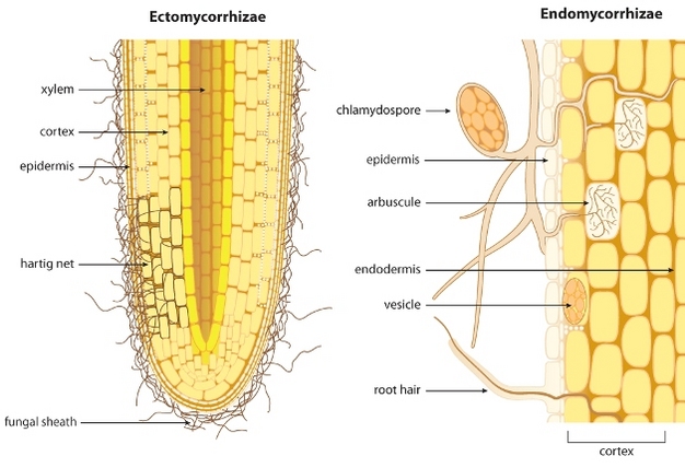
There are two broad categories of mycorrhizal associations with plant roots, ectomycorrhiza and endomycorrhiza, which are differentiated by how they physically interface with the plant (Figure 6). The ectomycorrhizae (EM) occur mainly in the roots of woody plants (i.e. forest trees) and form a dense hyphal covering (fungal sheath or mantel) over the root tip from which hyphae grow into the intercellular spaces forming a net (Hartig net) of hyphae around the root cortex cells, but do not penetrate the cell walls. In contrast, the endomycorrhizae fungal hyphae grow into the root cortex and enter the cells forming fan-like, highly branched structure known as an arbuscule that remain separated from the cytoplasm by the plant plasma membrane (Harrison, 2005). The endomycorrhiza can be further divided into the more widespread arbuscular mycorrhiza (AM) and the specialized orchid and ericoid mycorrhizas which, as the name implies, are colonizers of orchids and ericoid (e.g., cranberry) plant species. The AM fungal associations are the most abundant of all mycorrhizal associations. In both cases, the Hartig net and the arbuscules increase the contact area between the fungus and the plant through which the transfer of nutrients to the plant and carbon to the fungus occurs. Unlike the ectomycorrhiza, the endomycorrhiza are wholly dependent on the plant for their carbon and when associations occur, both endomycorrhiza and ectomycorrhiza can demand up to 20-40% of the total photosynthetically fixed carbon the plant produces.
Schematic showing the difference between ectomycorrhizae and endomycorrhizae

Figure 6
Schematic showing the difference between ectomycorrhizae and endomycorrhizae colonization of plant roots.
© 2013 Nature Education Bonfante, P. & Genre, A. Mechanisms underlying beneficial plant-fungus interactions in mycorrhizal symbiosis. Nature communications 1 doi:10.1038/ncomms1046. All rights reserved. View Terms of Use


The chemical dialogue that initiates mycorrhizal associations with plants is intricate and not as well understood as that of the Rhizobia. In part, the lack of understanding rests in the fact that endomycorrhiza are obligate symbionts, meaning they cannot be grown independent of their plant hosts, which makes it difficult for scientists to study them in the lab under controlled conditions. For this reason, more is known about the development of the ectomycorrhizal symbiosis because the two partners can be grown independently. In both cases, there is evidence that the fungus can sense the presence of volatiles (CO2), or chemicals in plant root exudates which then initiates hyphal growth and branching (Martin et al., 2001). Conversely, chemical signals from mycorrhizea which are perceived by the plant have long been suspected (Kosuta et al., 2003) and only recently has the structure of one of these chemicals in the ecotmycorrhizal association been fully characterized (Maillet et al., 2011). The chemical signals are aptly called Myc-factors which, interestingly, are similar in structure to the nod factors produced by rhizobia in the rhizobia-legume symbiosis. It is still unclear whether the fungus produces the signals all the time or only when it senses the plant is near. Regardless, fungal spores in the soil remain in a resting state until one of these stimuli initiates germination and hyphal growth. If close to a root, hyphal growth and branching will increase and Myc-factors will initiate root branching presumably to improve the odds of root-fungus interception (Figure 5b). After making contact with the plant root, infection begins by either the formation of a hyphopodium (in arbuscular mycorrhizae) or growth between dermal cells (in ectomycorrhizea). If a root is not in the vicinity, the hyphae stops growing and the spore returns to a vegetative state; subsisting off of its triacylglyceride and glycogen reserves. Initiation of hyphal growth can occur multiple times until root interception is achieved. To capture nutrients for the plant, both the ecto- and endomycorrhizae extend hyphae centimeters into the soil resulting in a 10 fold increase in the effective root surface area and a 2-3 fold increase in the uptake of phosphorus (and other nutrients) per unit root length compared to non-mycorrhizal plants. It is not only the amount of hyphae that helps in nutrient acquisition, but also their small size (<200 mm) that enables them to access small soil pores and cracks that the plant root would otherwise not be able to access. The network of fungal hyphae emanating from the plant roots also has a tremendous impact on soil quality. The mycorrhizal hyphae promote soil aggregate formation and stability via biological, physical and biochemical mechanisms which reduces soil erosion and increases soil aeration and water infiltration, which together improves plant productivity (Rillig and Mummey, 2006). Interestingly, the dense, intertwined network of fungal hyphae forms a "common mycorrhizal network" (CMN), in which hyphae from mycorrhizae infecting two or more plants are interconnected. Through the CMN, plants have been shown to share nutrients and mediate the interactions between plants which are not immediately sharing the same space (Selosse, el. al., 2006). The diversity of these interactions and the mycorrhizal fungus taking part in them have been shown to have a great deal of influences on plant biodiversity, ecosystem function and stability (van der Heijden, et al., 1998).


Plant Growth Promoting Rhizobacteria (PGPR)​


Plant growth promoting rhizobacteria were first defined by Kloepper and Schroth (1978) as organisms that, after being inoculated on seeds, could successfully colonize plant roots and positively enhance plant growth. To date, there have been over two dozen genera of nonpathogenic rhizobacteria identified. Plant growth promotion can be shown to work directly on the plant in the absence of root pathogens by the release of plant growth stimulating compounds (e.g. phytohormones such as auxins or cytokinins) and improvement in mineral uptake (e.g. siderophore release increasing Fe availability). Plant growth promotion can also occur indirectly by control of pathogens (biocontrol) via synthesis of antibiotics or secondary metabolite-mediated induced systemic resistance (ISR) (van Loon, et al., 1998, 2007).
Microbial colonization of the plant root surface is not uniform, but instead occurs in patches along the root, ultimately covering ~15- 40% of the total plant root surface. The density and structure of the microbes on the root surface are dictated by nutrient availability and physicochemical variations throughout the root surface. Root exudates can serve as a food source and chemoattractant for microbes which then attach to the root surface and form microcolonies. Common sites for bacterial attachment and colonization are at epidermal cell junctions, root hairs, axial groves, cap cells, and sites of emerging lateral roots (Danhorn et al., 2007). Microcolonies can eventually grow into larger biofilms consisting of multiple layers of bacteria which are encased in an exopolymeric matrix (figure 5c). In many cases the effectiveness of rhizobacteria at promoting growth occurs in a density dependant manner (Rudrappa, et al., 2008). Once a critical microbial density is reached, the biofilms then begin to act in unison, in a process known as quorum sensing, coordinating the release of compounds that aid in the promotion of plant growth via the direct and indirect mechanism discussed previously.

Root System Architecture — Sensing and Foraging​

The spatial extent and influence of the rhizosphere are dictated by root system architecture (RSA) (Figure 2). Root system architecture is very malleable, determined by plant species and occurring in response to changing climactic, biological and edaphic conditions. The distribution of nutrients in soils is heterogeneous or patchy and there is evidence plants can "sense" the presence of nutrients and allocate more resources to the root system and direct root growth toward or within these patches (Figure 7). Examples include increased root depth in drought tolerant beans, wheat, and maize and an increase of dense shallow root systems leading to topsoil foraging for P in plants under low P conditions (Ho et al, 2005). The nutrients themselves and the activity of specific molecular pathways involved in their acquisition may act to induce hormonal signals triggering increases in root density and length (Lopez-Bucio et al., 2003). Because of the underground nature of plant roots, uncovering the mechanisms responsible for root foraging has been difficult, but full annotation of the Arabidopsis genome and a complete map of root cell types have dramatically increased our understanding of root biology and these processes (Malamy and Benfey, 1997; Dinneny, et al, 2008; Benfey et al, 2010).
Changes in roots system architecture

Figure 7
Changes in roots system architecture (RSA) of barely (Hordeum vulgare) in response to zones of high phosphate, nitrate, ammonium and potassium availability.
© 2012 Nature Education Hodge, A. The plastic plant: root responses to heterogeneous supplies of nutrients. New Phytol 162, 9–24 (2004). All rights reserved. View Terms of Use


Summary​

Roots serve many functions for a plant including anchorage and acquisition of vital nutrients and water necessary for growth. The plant root-soil interface is a dynamic region in which numerous biogeochemical processes take place driven by the physical activity, and the diversity of chemicals released by the plant root and mediated by soil microorganisms. In turn the processes occurring in this region control a host of reactions regulating terrestrial carbon and other element cycling that sustain plant growth and which have an enormous influence on plant and microbial community function and structure which greatly influence a variety of ecosystem level processes (van der Heijden et. al., 2008; Wardle, 2004; Berg and Smalla, 2009). Understanding and harnessing these interactions for the sustainable production of food, fuel and fiber to support a growing world population on a dwindling supply of arable land will be the challenge of generations to come.


Glossary​


arbuscule - a highly branched fungal structure occurring within the cortical cells of roots colonized by arbuscular mycorrhizal fungi (Sylvia et al. 2005)
biocontrol - the use of microorganisms to suppress diseases and improve overall plant health (Handelsman and Stabb 1996)
biofilms -assemblage of many microbial cells bound together by an adhesive substance (often polysaccharides) and attached to a surface (Sylvia et al. 2005)
chelate -a molecule with multiple binding sites that attaches to a metal ion (Petrucci 1989)
chemoattractant - compounds released by plants or microorganisms which attract microbes to the plant or direct their movement often towards one another (Park et al. 2003)
clay minerals - the chemical weathering products of rocks, composed principally of parallel silica sheets (phyllosilicates), that impart plasticity to soils and harden when dry.
cortex - the tissue of a root confined externally by the epidermis (the "skin" of the root) and internally by the endodermis (the tissues encircling the vascular tissues) (Raven et al. 1999)
denitrification - the process whereby nitrate or nitrite is reduced to N2 (g) primarily by microorganisms proceeding as follows: NO3- → NO2- → NO + N2O → N2 (g) (Sylvia et al. 2005)
dicot - One of the two main divisions (the other being monocots) of flowering plants (i.e. angiosperms) which have two cotyledons or "seed leaves". Examples include fruiting trees and most annual and perennial flowering plants.
diffusates - a subset of exudates which are passively released from the root (Pinton et al. 2007)
ectorhizosphere - the area surrounding (extracellularly) the root where microbes fed by root derived compounds may colonize (El-Morsy 1999; Lynch 1982)
ectomycorrhizae - a plant-fungus relationship where the fungal hyphae extend into the plant root and occupy the area between the cortical cells, forming a Hartig net (Sylvia et al. 2005)
edaphic - of, pertaining to, or influenced by the soil (Sylvia et al. 2005)
endodermis - a single layer of cells enclosing the vascular tissue of the root (Raven et al. 1999)
endomycorrhizae - a plant-fungus relationship where the fungal hyphae extend into the plant root and occupy the area between and within (intracellularly) the cortical cells (Sylvia et al. 2005)
endorhizosphere - the region in the root which microbes fed by root derived compounds may colonize including root cortex, epidermis and root hairs (Kloepper et al. 1992a; Lynch 1982)
exudates - compounds released into the soil/rhizosphere by plant roots (Walker et al. 2003)
hartig net - a net-like structure created by ectomycorrhizae fungal hyphae which envelops the cortical cells of the plant root and facilitates nutrient exchange between the plant and the fungal host (Sylvia et al. 2005)
hyphopodium -Specialized hyphal branch composed of one or two swollen hyphal branches serving for attachment to the root epidermis and absorption of nutrients (Genre, 2007)
induced systemic resistance (ISR) - the protection of the whole plant induced by an external agent applied to a localized region (Liu et al. 1995; Kloepper et al. 1992b)
mucilage - a high molecular weight mixture of various polysaccharides (Knee et al. 2001) primarily secreted from the root cap and border cells (Hawes et al. 1998)
monocot - One of the two (the other being dicots) divisions of flowering plants (i.e. angiosperms) which have only one cotyledon ("seed leaf") and parallel veins. The most common examples are grasses like corn, barley and wheat.
mycorrhizae - a term used to describe a symbiotic association between root colonizing fungi and plants (Sylvia et al. 2005)
obligate symbionts - organisms which rely on a host organism to provide the carbon and other nutrients required for life (Sylvia et al. 2005)
phytohormones - plant derived hormones which regulate physiological processes (Raven et al. 1999)
phytosiderophores - low molecular weight compounds (e.g., mugienic acid) released from plant roots which chelate Fe, thereby, making it more available for the plant (Buchanan et al. 2001)
quorum sensing - a phenomenon whereby the gene regulation of an individual microbial cell is influenced in a cell-population density dependent manner through the release of signaling chemicals which increase as the population grows (Miller and Bassler 2001)
redox - oxidation-reduction reactions are coupled reactions whereby one compound releases electrons (becoming oxidized) while another compound absorbs the electrons becoming reduced (Sylvia et al. 2005). The term "redox condition" refers to the environmental parameters controlling redox reactions (i.e. oxygen concentration, pH, temperature, and availability of terminal electron acceptors).
rhizodeposits - root derived carbon sources imparted to soils from growing plants (Pinton et al. 2007)
rhizoplane - the root surface including associated soil particles (Estermann and McLaren 1961; Clark 1949)
rhizosphere - the zone of chemical, biological, and physical influence generated by root growth and activity. The concept usually pertains to the soil-root interface but is sometimes extrapolated to other media-root interfaces (Pinton et al. 2007)
secretions - a subset of exudates which are actively released from the root (Pinton et al. 2007)
siderophore - (Greek for "iron carrier") compounds released from bacteria, fungi and grasses with an extremely high affinity for iron (Leong and Neilands 1976)
strategy I - a mechanism of Fe acquisition whereby H+ is released from the roots and increases the amount of free Fe in solution (Römheld and Marschner 1986)
strategy II - a mechanism of Fe acquisition whereby phytosiderophores (Fe chelating compounds) are released and lead to increased plant available Fe (Römheld and Marschner 1986)


References and Recommended Reading​


Bais, H. P., Park, S. W. , Weir, T.L., Callaway, R.M. and Vivanco, J.M. How plants communicate using the underground information superhighway. Trends in Plant Science 9(1):26-32. (2004)
Benfey, P.N., Bennett, M., Scheifelbein, J. Getting to the root of plant biology: impact of the Arabidopsis genome sequence on root research. The plant journal. 61:992-1000. (2010).
Bengough, A.G. and McKenzie, B.M. Sloughing of root cap cells decreases the frictional resistance to maize (Zea mays L.) root growth. J. Exper. Bot, Vol. 48(309):885-893 (1997)
Berg, G. Smalla, K. Plant species and soil type cooperatively shape the sturcutre and function of microbial communities in the rhizosphere. FEMS Microbiol. Ecol. 68:1-13 (2009)
Buchanan, B. B., Gruissem, W. & Jones, R. L. Biochemistry and Molecular Biology of Plants (Courier Companies Inc., U.S., 2001).
Clark, F. E. Soil Micro-organisms and Plant Roots (1949).
Danhorn T, Fuqua C. Biofilm formation by plant-associated bacteria. ANNUAL REVIEW OF MICROBIOLOGY. 61: 401-422 ( 2007)
Dinneny JR, Long TA, Wang JY, Jung JW, Mace D, Pointer S, Barron C, Brady SM, Schiefelbein J, Benfey PN. Cell Identity Mediates the Response of Arabidopsis Roots to Abiotic Stress. (2008) Science.
El-Morsy, E. M. Microfungi from the ectorhizosphere-rhizoplane zone of different halophytic plants from the Red Sea coast of Egypt. Mycologia 91: 228-236 (1999).
Estermann, E. F. & McLaren, A. D. Contribution of rhizoplane organisms to the total capacity of plants to utilize organic nutrients. Plant and Soil 15, 243-260 (1961).
Genre, A. and P. Bonfante (2007). "Check-In Procedures for Plant Cell Entry by Biotrophic Microbes." Molecular Plant Microbe Interactions 20(9): 1023-1030.
Gewin, V. Food: An underground revolution. Nature 466: 552-553 (2010)
Giles E.D. Oldroyd and J. Allan Downie. Coordinating Nodule Morphogenesis with Rhizobial Infection in Legumes. Annual Review of Plant Biology. 59: 519-546 (2008)
Handelsman, J. & Stabb, E. V. Biocontrol of soilborne plant pathogens. Plant Cell 8, 1855-1869 (1996).
Harrison M.J. Signaling in the arbuscular mycorrhizal symbiosis. Annu. Rev. Microbiol. 59:19-24 (2005)
Hartmann A., Rothballer M., and M. Schmid. Lorenz Hiltner, a pioneer in rhizosphere microbial ecology and soil bacteriology research. Plant Soil 312:7-14 (2008)
Hawes, M. C., Brigham, L. A., Wen, F., Woo, H. H. & Zhu, Z. Function of root border cells in plant health: Pioneers in the rhizosphere. Annual Review of Phytopathology 36, 311-327 (1998).
Hawes MC, Gunawardena U, Miyasaka S, et al. The role of root border cells in plant defense. TRENDS IN PLANT SCIENCE 5(3):128-133 (2000)
Hellriegel H, Wilfarth H Untersuchungen uber die Stickstoffnahrung der Gramineon und Leguminosen. Beilageheft zu der Ztschr. Ver. Ru¨ benzucker-Industrie Deutschen Reichs (1888)
HILTNER, L. Ueber neuere Erfahrungen und Probleme auf dem Gebiete der Bodenbakteriologie und unter besonderer BerUcksichtigung der Grundungung und Brache. Arb. Deut. Landw. Gesell, 98:59-78. (1904)
Hirsch, A.M., Lum, M.R. and Downie, J.A. What makes the rhizobia-legume symbiosis so special? Plant. Phys. 127:1484-1492. (2001)
Ho, M.D., Rosas, J.C., Brown, K.M., Lynch, J.P. Root architecture tradeoffs from water and phosphorus acquisition. Functional Plant Biology. 32:737-748. (2005)
Hodge, A. (2004) The plastic plant: root responses to heterogeneous supplies of nutrients. New Phytol. 162, 9-24
Jones, K.M., Kobayashi, J., Davies, B.W., Taga, M.E., and Walker, G.C. How rhizobial symbionts invade plants: the Sinorhizobium-Medicago model. Nature Reviews Microbiology. 5(8):619-633. 2007
Jones, D.L., Nguyen, C., and Finlay, R.D. Carbon flow in the rhizosphere: carbon trading at the soil-root interface. Plant Soil. 321:5-33 (2009).
Kloepper, J. W. & Schroth, M. N. in Proc 4th int. Conf. Plant Pathogenic Bacteria Vol. 2 (ed. Station de Pathologie Végétale et Phytobactériologie) 879-882 (Gibert-Clarey, Tours, 1978)
Kloepper, J. W., Schippers, B. & Bakker, P. A. H. M. Proposed Elimination of the Term Endorhizosphere. Phytopathology 82, 726-727 (1992a).
Kloepper, J. W., Tuzun, S. & Kuc, J. A. Proposed Definitions Related to Induced Disease Resistance. Biocontrol Science and Technology 2, 349-351 (1992b).
Knee, E. M. et al. Root mucilage from pea and its utilization by rhizosphere bacteria as a sole carbon source. Molecular Plant-Microbe Interactions 14, 775-784 (2001).
Kosuta, S., Chabaud, M., Lougnon, G., Gough, C., Denarie, J., Barker, D.G., Becard, G. A diffusible factor from arbuscular mycorrhizal fungi induces symbiosis-specific MtENOED11 expression in roots of Medicago truncatula. Plant Physiol. 131:952-962 (2003).
Leong, J. & Neilands, J. B. Mechanisms of siderophore iron transport in enteric bacteria. J. Bacteriol. 126, 823-830 (1976).
Liu, L., Kloepper, J. W. & Tuzun, S. Induction of Systemic Resistance in Cucumber by Plant Growth-Promoting Rhizobacteria - Duration of Protection and Effect of Host-Resistance on Protection and Root Colonization. Phytopathology 85, 1064-1068 (1995).
Lopez-Bucio J, Cruz-Ramirez A, Herrera-Estrella L. The role of nutrient availability in regulating root architecture. CURRENT OPINION IN PLANT BIOLOGY 6(3): 280-287 (2003)
Lynch, J. in Experimental Microbial Ecology (eds. Burns, R. G. & Slater, J. H.) (Blackwell Scientific Publications 1982).
Maillet, F., Poinsot, V.,Andre, O., Puech-Pages, V., Haouy, A., Gueunier, M., Cromer, L., Giraudet, D., Formey, D., Niebel, A., Andres Martinez, E., Driguez, H., Cecard, G., Denarie, J. Fungal lipochitooligosaccharide symbiotic signals in arbuscular mycorrhizal. Nature. 469:58-64 (2011)
Malamy, J.E. and Benfey, P.N. Organization and cell differentiation in laterl roots of Arabidopsis thaliana. Development. 124:33-44. (1997)
Martin, F., Duplessis, S., Ditengou, F., Lagrange, H., Voiblet, C., and Lapeyrie, F. Developmental cross talking in the ectomycorrhizal symbiosis: signals and communication genes. New Phytologist. 151:145-154. (2001)
Miller, M. B. & Bassler, B. L. Quorum Sensing in Bacteria. Annual Review of Microbiology 55, 165-199 (2001).
Morrisseey, J., Dow, J., Mark, G., O'Gara, F. Are microbes at the root of a solution to world food production? EMBO rept. 5(10) 922-926. (2004)
Newman, E. I. The rhizosphere: carbon sources and microbial populations. Ecological Interactions in Soil. A. H. Fitter. Oxford, Blackwell Scientific Publications: p. 107. (1985)
Park, S. et al. Motion to Form a Quorum. Science 301, 188 (2003).
Petrucci, R. H. General Chemistry (Macmillan Publishing Company, New York, New York, 1989).
Pinton, R., Varanini, Z. & Paolo, N. The Rhizosphere: Biochemistry and Organic Substances at the Soil-Root Interface (CRC Press, Boca Raton, 2007).
Raghothama, K.G. Phosphate acquisition. Annu. Rev. Plant Physiol. Plant Mol. Biol. 50:665-693. (1999)
Raven, P. H., Evert, R. F. & Eichhorn, S. E. Biology of Plants (W.H. Freeman and Company Worth Publishers, New York, New York, 1999).
Rillig M.C., and D.L. Mummey. Mycorrhizas and soil structure. New Phytologist, 171:41-53. (2006)
Römheld, V. & Marschner, H. Evidence for a Specific Uptake System for Iron Phytosiderophores in Roots of Grasses. Plant Physiology 80, 175-180 (1986).
Rovira, A.D. Plant root exudates. Botanical Review. 35(1):35-57 (1969)
Rudrappa, T., Biedrzycki, M.L. and Bais, H.P. Causes and consequences of plant-assocaited biofilms. FEMS Mirobial. Ecol. 64:153-166. (2008)
Selosse, M.A., Richard, F., He, X., Simard, S.W. Mycorrhizal networks" des liaisons dangereuses. TRENDS Ecol. Evol. 21(11):621-628. (2006)
Sylvia, D. M., Fuhrmann, J. J., Hartel, P. G. & Zuberer, D. A. Principles and Applications of Soil Microbiology (Pearson Prentice Hall, Upper Saddle River, New Jersey, 2005).
Tilman, D., Cassman, K. G., Matson, P. A., Naylor, R. & Polasky, S. Agricultural sustainability and intensive production practices. Nature. 418, 671-677 (2002).
van der Heijden, M.G.A., Bardgett, R.D., and vanStraalen, N.M. The unseen majority" soil microbes as drivers of plant diversity and productivity in terrestrial ecosystems. Ecology Letters. 11:296-310 (2008)
van der Heijden, M.G.A., Klironomos JN, Ursic M, Moutoglis P, Streitwolf-Engel R, Boller T, Wiemken A, Sanders IR. Mycorrhizal fungal diversity determines plant biodiversity, ecosystem variability and productivity. Nature. 396(6706): 69-72 (1998)
van Loon, L.C., Bakker, P. A. H. M. and Pieterse, C. M. J. SYSTEMIC RESISTANCE INDUCED BY RHIZOSPHERE BACTERIA. Annual Review of Phytopathology. 36: 453-483 (1998)
van Loon, L.C. Plant responses to plant growth-promoting rhizobacteria. Eur. J. Plant Pathol. 119:243-254. (2007).
Walker, T. S., Bais, H. P., Grotewold, E. & Vivanco, J. M. Root Exudation and Rhizosphere Biology. Plant Physiology 132, 44-51 (2003).
Wardle DA, Bardgett RD, Klironomos JN, Setala H, van der Putten WH, Wall DH. Ecological linkages between aboveground and belowground biota. SCIENCE. 304(5677): 1629-1633 (2004)
Wilcox HE. 1991. Mycorrhizae. In: Waisel Y, Eshel A, Kafkaki U, eds. Plant roots: the hidden half. New York, USA: Marcel Dekker
Zhang, F., Shen, J., Zhang, J., Zuo, Y. Li, L., Chen, X. Rhizosphere processes and management for improving nutrient use efficiency and crop productivity: implications for China. Advances in Agronomy. 107:1-32 (2010)
 

acespicoli

Well-known member
Polymers (Basel). 2023 Jul; 15(13): 2867.
Published online 2023 Jun 28. doi: 10.3390/polym15132867
PMCID: PMC10346603
PMID: 37447512

Chitosan: Properties and Its Application in Agriculture in Context of Molecular Weight​

Ramón Román-Doval,1,* Sandra P. Torres-Arellanes,1 Aldo Y. Tenorio-Barajas,2 Alejandro Gómez-Sánchez,1 and Anai A. Valencia-Lazcano3,*
Balzhima Shagdarova, Academic Editor and Alla Il’ina, Academic Editor

1721232674668.png

The cheapest way to lower the soil pH is to add elemental sulfur to the soil.
Soil bacteria change the sulfur to sulfuric acid,

Uptake via plant roots

Once this conversion has occurred, the plant can absorb the sulfate through its roots. Inside the plant, the sulfate is a key ingredient in many key plant processes: from the production of chlorophyll to the synthesis of starches, sugars oils, fats and vitamins.

Slow-release for optimal results

It’s important to note that microorganisms cannot oxidize elemental sulfur overnight. The process can take weeks and is affected by many factors (such as moisture, soil conditions, temperature, microbe populations and fertilizer quality).

Because this conversion process takes time, elemental sulfur is considered a slow-release fertilizer. Fall-applying elemental sulfur gives the microbes a head start, allowing the conversion process to begin sooner, resulting in a ready supply of sulfate to seedlings from the outset of the crop year.

Creating the right conditions

Having a healthy population of the right microbes is necessary for the conversion from S to SO4 to take place. Inoculation has not proven to be an effective solution for increasing populations of Thiobacillus


Dictionary
Definitions from Oxford Languages · Learn more


thio-
/ˈTHīō/
combining form
CHEMISTRY

  1. denoting replacement of oxygen by sulfur in a compound.
https://en.wikipedia.org/wiki/Thiobacillus
and other microorganisms responsible for sulfur oxidation.

However, the presence of sulfur will stimulate population growth by providing conditions where they can thrive.

Other factors that can impact the efficacy of elemental sulfur include:

  • Soil moisture: Moisture is required to activate the bentonite clay to swell, and break down sulfur particles to the optimum size required for conversion.
  • Soil temperature: Optimum soil conditions for oxidization range between 75-105°F (24-40°C).
  • Soil pH: Thiobacillus populations thrive in acidic soils, which also promote the speed of oxidization.
  • Soil health: Microorganisms, like all living creatures, require a variety of nutrients to survive.
The bottom line? Your bentonite sulfur fertilizer can go a long way to not only giving your crop the best conditions for success but also supporting sustainable high-quality soils for a great harvest year after year, all thanks to a wonderful symbiotic relationship with a healthy microbial population
 
Last edited:

acespicoli

Well-known member
1721234842706.png


Soil beneficial bacteria and their role in plant growth promotion: a review​

Annals of Microbiology volume 60, pages579–598 (2010)Cite this article
Ann Microbiol (2010) 60:579–598
DOI 10.1007/s13213-010-0117-1


There are several dozen recognized subspecies of B. thuringiensis. Subspecies commonly used as insecticides include B. thuringiensis subspecies kurstaki (Btk), subspecies israelensis (Bti) and subspecies aizawai (Bta).[19][20][21][22] Some Bti lineages are clonal.[18]

Common disease resistance mechanisms[edit]​

Pre-formed structures and compounds[edit]​

secondary plant wall

Inducible post-infection plant defenses[edit]​


Solubility in a strong or weak acid solution[edit]​

Solutions of strong (HCl), moderately strong (sulfamic) or weak (acetic, citric, sorbic, lactic, phosphoric) acids are commercially available. They are commonly used as descaling agents to remove limescale deposits. The maximum amount of CaCO3 that can be "dissolved" by one liter of an acid solution can be calculated using the above equilibrium equations.

  • In the case of a strong monoacid with decreasing acid concentration [A] = [A−], we obtain (with CaCO3 molar mass = 100 g/mol):
[A] (mol/L)110−110−210−310−410−510−610−710−10
Initial pH0.001.002.003.004.005.006.006.797.00
Final pH6.757.257.758.148.258.268.268.268.27
Dissolved CaCO3 (g/L of acid)50.05.000.5140.08490.05040.04740.04710.04700.0470
where the initial state is the acid solution with no Ca2+ (not taking into account possible CO2 dissolution) and the final state is the solution with saturated Ca2+. For strong acid concentrations, all species have a negligible concentration in the final state with respect to Ca2+ and A− so that the neutrality equation reduces approximately to 2[Ca2+] = [A−] yielding [Ca2+] ≈ 0.5 [A−]. When the concentration decreases, [HCO−3] becomes non-negligible so that the preceding expression is no longer valid. For vanishing acid concentrations, one can recover the final pH and the solubility of CaCO3 in pure water.
  • In the case of a weak monoacid (here we take acetic acid with pKa = 4.76) with decreasing total acid concentration [A] = [A−] + [AH], we obtain:
 
Last edited:

acespicoli

Well-known member
Microbiology (Reading). 2021; 167(4): 001049.
Published online 2021 Apr 21. doi: 10.1099/mic.0.001049
PMCID: PMC8289221
PMID: 33881981

Bacteria-induced mineral precipitation: a mechanistic review​

Timothy D. Hoffmann, 1 ,* Bianca J. Reeksting, 1 and Susanne Gebhard 1 ,*
Author information Article notes Copyright and License information PMC Disclaimer

Go to:

Abstract​

Micro-organisms contribute to Earth’s mineral deposits through a process known as bacteria-induced mineral precipitation (BIMP). It is a complex phenomenon that can occur as a result of a variety of physiological activities that influence the supersaturation state and nucleation catalysis of mineral precipitation in the environment. There is a good understanding of BIMP induced by bacterial metabolism through the control of metal redox states and enzyme-mediated reactions such as ureolysis. However, other forms of BIMP often cannot be attributed to a single pathway but rather appear to be a passive result of bacterial activity, where minerals form as a result of metabolic by-products and surface interactions within the surrounding environment. BIMP from such processes has formed the basis of many new innovative biotechnologies, such as soil consolidation, heavy metal remediation, restoration of historic buildings and even self-healing concrete. However, these applications to date have primarily incorporated BIMP-capable bacteria sampled from the environment, while detailed investigations of the underpinning mechanisms have been lagging behind. This review covers our current mechanistic understanding of bacterial activities that indirectly influence BIMP and highlights the complexity and connectivity between the different cellular and metabolic processes involved. Ultimately, detailed insights will facilitate the rational design of application-specific BIMP technologies and deepen our understanding of how bacteria are shaping our world.
Keywords: biomineralization, organomineralization, biologically induced mineralization, nucleation, biogenic
Go to:

Introduction​

Bacterial activity is evident in our landscapes and throughout the geological record, where it has helped shape Earth’s mineral deposits [1]. This has occurred, to some degree, via a process known as bacteria-induced mineral precipitation (BIMP). The variety of mineral deposits that are formed through bacterial activity can take on the form of stalactites and stalagmites [2], microbialites, stromatolites and thrombolites [3, 4] as well as large-scale sedimentation [4]. More recently, the ability of bacteria to induce mineral formation has gained attention for biotechnological application. In particular, the precipitation of calcium carbonate in the form of calcite, the mineral that forms limestone, has been exploited in innovative technologies in civil engineering. The first patented application is considered to have been by Adolphe and colleagues in 1990 for biological treatment of degrading stone surfaces [5]. Since then, more technologies have been developed, with a lot of attention surrounding the concept of self-healing concrete [6–8]. Other applications of BIMP include soil consolidation or heavy metal bioremediation, and excellent recent reviews exist that cover the spectrum of such technologies in detail [9–14].
For the purposes of this review, BIMP is defined as a process by which bacterial activity indirectly induces mineral formation via the release of metabolic by-products and surface interactions with ions in the open environment [15–17]. This is in contrast to bacteria-controlled biomineralization, e.g. the formation of magnetite by magnetotactic bacteria, which is metabolically and genetically controlled by the bacteria and occurs in defined locations, e.g. magnetosomes [18–20]. The latter has been reviewed in detail elsewhere [15, 21] and will not be covered here. The minerals formed by BIMP generally have no specific function (aside from some potential ecological benefits) and can be considered an unintended and uncontrolled consequence of bacterial activity [22, 23]. Depending on the author, indirect biomineralization is sometimes subdivided further into more nuanced ‘bacteria-induced’ versus ‘bacteria-influenced’ mineral precipitation [13, 20, 24]. The boundaries between the two are, however, not clear cut and in this review no such division is made.

Bacteria-induced mineral precipitation​

Precipitation of mineral species in an aqueous system occurs when the ion concentration exceeds solubility and reaches a degree of super-saturation. Once the activation energy barrier is overcome, initial crystal nucleation occurs, in which metastable critical nuclei form that may dissolve back into the bulk phase. Subsequent aggregation of individual nuclei describes the process of crystal growth and precipitation [25–27]. Nucleation can take place either homogeneously, whereby nucleation occurs when critical nuclei form in the absence of foreign particles (via random collisions of ions or atoms in solution), or heterogeneously, whereby nucleation takes place when critical nuclei form on surfaces of foreign particles [25–27]. Such particles lower the activation energy by providing templates with spacing that enhances nucleation and thus, precipitation [25–27]. Furthermore, during the nucleation process foreign particles may aggregate, leading to the formation of mixed precipitates [28].
In BIMP, bacteria can induce biomineralization by modulating precipitation-relevant parameters like local ion concentrations or pH in the environment and/or by bacterial cells themselves providing nucleation sites for crystal formation. In general, this bacterial process involves the attraction of cations to negative charges on the cell surfaces, while metabolic activity provides the appropriate microenvironment and counter-anions so that these cations may precipitate as minerals [29]. The BIMP trait is common amongst bacteria across environments [9, 30–33], and, depending on bacterial species and environment, it can lead to a range of precipitated minerals (Table 1). The bacteria-induced formation of some of these minerals can further lead to co-precipitation of additional divalent metal cations and anions [34–36]. Indirect bacterial influence on precipitation parameters of saturation state and nucleation catalysis can be broadly separated into two contributing areas: cell surface and metabolic activity, and our current understanding of the mechanisms of these will be reviewed here.

Table 1.​

Minerals precipitated in association with bacterial activity*
Mineral​
Chemical formula​
Reference​
Carbonates
Calcite​
CaCO3​
[30]​
Dolomite​
CaMg(CO3)2​
Kutnahorite​
CaMn(CO3)2​
[113]​
Siderite​
FeCO3​
[114]​
Magnesite​
MgCO3​
[54, 115]​
Otavite​
CdCO3​
[116]​
Strontianite​
SrCO3​
[72]​
Rhodochrosite​
MnCO3​
[117]​
Cerussite​
PbCO3​
[118]​
Hydrozincite​
Zn5(CO3)2(OH)6​
[36, 119]​
Dypingite​
Mg5(CO3)(OH)2·5H2O​
[120]​
Witherite​
BaCO3​
[121]​
Phosphates
Tricalcium phosphate​
Ca3(PO4)2​
[78]​
Struvite​
NH4MgPO4∙6H2O​
[74, 113]​
Bobierrite​
Mg3(PO4)2·8H2O​
[74, 122]​
Baricite​
(MgFe)3(PO4)2·8H2O​
[74]​
Vivianite​
Fe3(PO4)·2H2O​
[114]​
Autunite​
Ca(UO2)2(PO4)2∙10-12H2O​
[44]​
Uramphite​
NH4UO2PO4​
Apatite​
Ca10(PO4)6(OH)2​
[124]​
Pb-hydroxyapatite​
Ca2.5Pb7.5(OH)2(PO4)6​
[125]​
Strengite​
FePO4·2H2O​
Variscite​
AlPO4·2H2O​
[97]​
Silicates
Gehlenite​
Ca2Al(AlSiO7)​
[128]​
Silica​
SiO2​
[129]​
Nontronite​
Na0.3Fe3+ 2(Si,Al)4O10(OH)2·nH2O​
[130]​
Chamosite​
(Fe5Al)(Si3Al)10(OH)8​
[126]​
Kaolinite​
Al4(Si4O10)(OH)4​
[126]​
Sulphides
Mackinawite​
FeS​
[76]​
Greigite​
Fe3S4​
[76, 131]​
Pyrite​
FeS2​
[132]​
Covellite​
CuS​
Sphalerite​
ZnS​
[135]​
Galena​
PbS​
[134]​
Digenite​
Cu9S5​
[136]​
Sulphates
Gypsum​
CaSO4·2H2O​
[41, 54]​
Celestite​
SrSO4​
[72]​
Barite​
BaSO4​
Oxides
Magnetite​
Fe3O4​
[114]​
Hematite​
Fe2O3​
[23, 138]​
Ferrihydrite​
Fe2O3·0.5H2O​
[138]​
Geothite​
α-FeO(OH)​
[138]​
Manganite​
MnOOH​
[139]​
Vernadite​
MnO2​
Hausmannite​
Mn3O4​
[142]​
Todorokite​
(Ca,Na,K)x(Mn4+,Mn3+)6O10·3.5H2O​
[138]​
Birnessite​
(Na,Ca,K)x(Mn4+,Mn3+)2O4·1.5H2O​
[138]​
Uraninite​
UO2​
Calcium Arsenate​
CaHAsO3​
[146]​
Open in a separate window
*Note that while these minerals have all been reported to be formed in association with bacterial activity, the mechanisms for their formation are not always known, and some minerals can be formed by multiple different mechanisms. The minerals listed and accompanying sources are non-exhaustive of the examples available in the literature.
 

acespicoli

Well-known member

Production of Calcite (Calcium Carbonate) Crystals by Soil Bacteria is a General Phenomenon​

Nature volume 246, pages527–529 (1973)Cite this article
Abstract
CERTAIN bacteria form crystals from the solutes in their aqueous environment, and some authors have associated this activity with the extensive deposits of CaCO3 in such places as the Grand Bahama, in spite of the belief that physicochemical effects, such as rapid changes in pH, salinity and temperature, are responsible1–4. Drew5 isolated a denitrifying bacterium able to form CaCO3 crystals in liquid media and named it “Bacterium calcis” (later named Pseudomonas calcis6). Greenfield7 obtained aragonite (another form of CaCO3) crystals in cultures of Pseudomonas in an artificial seawater medium containing Na2CO3 or (NH4)2CO3. Buck and Greenfield reported the same result with marine yeast, and claimed that calcium crystals resulted from the accumulation of calcium deposits on the surface of the cells8. McCallum and Guhathakurta9 observed calcium carbonate deposition by marine bacteria isolated from Bahama Island sediments, and when these were cultured in different media aragonite crystals formed. Shinano10 described many marine bacteria able to form crystals in liquid media. Ramos-Cormenzana11 has also reported crystal formation by soil bacteria in solid media. This, together with the knowledge that previous research concerned only marine bacteria in liquid media, stimulated us to investigate crystal formation by soil bacteria cultured on solid media.
 

acespicoli

Well-known member


Assignment to the elements in Kepler's Harmonices Mundi

Crystal Formation by Microorganisms in the Dunes and Soils at White Sands NM http://www.nps.gov/whsa/index.htm http://www.southwestlearning.org/ June 2012 Introduction Beneath your feet as you walk across the gypsum dunes and soils of White Sands National Monument is an ecosystem of roots and millions and millions of microorganisms that live in the pore spaces between sand grains. This invisible ecosystem is as complex as any rain forest on Earth. There are bacteria that capture, transform, and release nitrogen. There are complex networks of fungal hyphae that carry nutrients to roots. There are microscopic animals that feed on the fungi, and fungi that feed on the animals. Such subterranean ecosystems are some of the most populated and ancient ecosystems ever to exist on the planet. A handful of rich garden topsoil, for example, will contain more microorganisms than the seven billion people on Earth. Geologic evidence preserved in rocks reveals that microorganisms inhabited soils of ancient landscapes millions of years before plants colonized land. Although it is a widely known that organisms in the ocean produce calcium carbonate (calcite) as sea shells, it is less widely known that organisms in desert soils also produce calcite. The process of organisms making calcite, or any other mineral, is known as biomineralization. There are two types of biomineralization—“biologically controlled” and “biologically induced.” Biologically controlled occurs when organisms have specialized tissue where minerals are purposefully generated. Biologically induced occurs when organisms inadvertently create an extraneous environment that brings together the ingredients needed to make minerals. At White Sands National Monument, calcium carbonate appears to be biologically induced by many microorganisms, but it is unknown what type of microbes, how many, at what rate, and if the aboveground ecosystem is the main controlling factor for biomineralization.
 

acespicoli

Well-known member
The solution of these models has given insights into the nature of phase transitions, magnetization and scaling behaviour, as well as insights into the nature of quantum field theory.

Structure[edit]​

View of tetrahedral sheet structure of a clay mineral. Apical oxygen ions are tinted pink.
Like all phyllosilicates, clay minerals are characterised by two-dimensional sheets of corner-sharing SiO4 tetrahedra or AlO4 octahedra. The sheet units have the chemical composition (Al, Si)3O4. Each silica tetrahedron shares three of its vertex oxygen ions with other tetrahedra, forming a hexagonal array in two dimensions. The fourth oxygen ion is not shared with another tetrahedron and all of the tetrahedra "point" in the same direction; i.e. all of the unshared oxygen ions are on the same side of the sheet. These unshared oxygen ions are called apical oxygen ions.[20]

In clays, the tetrahedral sheets are always bonded to octahedral sheets formed from small cations, such as aluminum or magnesium, and coordinated by six oxygen atoms. The unshared vertex from the tetrahedral sheet also forms part of one side of the octahedral sheet, but an additional oxygen atom is located above the gap in the tetrahedral sheet at the center of the six tetrahedra. This oxygen atom is bonded to a hydrogen atom forming an OH group in the clay structure. Clays can be categorized depending on the way that tetrahedral and octahedral sheets are packaged into layers. If there is only one tetrahedral and one octahedral group in each layer the clay is known as a 1:1 clay. The alternative, known as a 2:1 clay, has two tetrahedral sheets with the unshared vertex of each sheet pointing towards each other and forming each side of the octahedral sheet.[20]

Bonding between the tetrahedral and octahedral sheets requires that the tetrahedral sheet becomes corrugated or twisted, causing ditrigonal distortion to the hexagonal array, and the octahedral sheet is flattened. This minimizes the overall bond-valence distortions of the crystallite.[20]

Depending on the composition of the tetrahedral and octahedral sheets, the layer will have no charge or will have a net negative charge. If the layers are charged this charge is balanced by interlayer cations such as Na+ or K+ or by a lone octahedral sheet. The interlayer may also contain water. The crystal structure is formed from a stack of layers interspaced with the interlayers.[20]

Classification[edit]​

Structure of clay mineral groups
Clay minerals can be classified as 1:1 or 2:1. A 1:1 clay would consist of one tetrahedral sheet and one octahedral sheet, and examples would be kaolinite and serpentinite. A 2:1 clay consists of an octahedral sheet sandwiched between two tetrahedral sheets, and examples are talc, vermiculite, and montmorillonite. The layers in 1:1 clays are uncharged and are bonded by hydrogen bonds between layers, but 2:1 layers have a net negative charge and may be bonded together either by individual cations (such as potassium in illite or sodium or calcium in smectites) or by positively charged octahedral sheets (as in chlorites).[9]
 
Last edited:

acespicoli

Well-known member
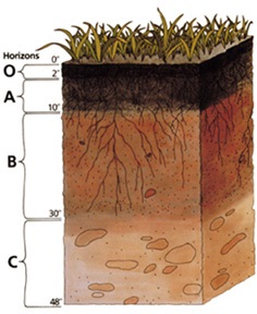
soil horizons

All soils have different types of layers.


There are different types of soil, each with its own set of characteristics. Dig down deep into any soil, and you’ll see that it is made of layers, or horizons

(O, A, E, B, C, R).

Put the horizons together, and they form a soil profile.
Like a biography, each profile tells a story about the life of a soil.
Most soils have three major horizons (A, B, C) and some have an organic horizon (O).

The horizons are:

O (humus or organic): Mostly organic matter such as decomposing leaves. The O horizon is thin in some soils, thick in others, and not present at all in others.

A (topsoil): Mostly minerals from parent material with organic matter incorporated. A good material for plants and other organisms to live.

E (eluviated): Leached of clay, minerals, and organic matter, leaving a concentration of sand and silt particles of quartz or other resistant materials – missing in some soils but often found in older soils and forest soils.

B (subsoil): Rich in minerals that leached (moved down) from the A or E horizons and accumulated here.

C (parent material): The deposit at Earth’s surface from which the soil developed.

R (bedrock): A mass of rock such as granite, basalt, quartzite, limestone or sandstone that forms the parent material for some soils

– if the bedrock is close enough to the surface to weather.

This is not soil and is located under the C horizon.
 

acespicoli

Well-known member
Soil Formation –

CLORPT – for short! Soils differ from one part of the world to another,
even from one part of a backyard to another.
They differ because of where and how they formed.
Five major factors interact to create different types of soils:

CLimate—Temperature and moisture influence the speed of chemical reactions, which in turn help control how fast rocks weather and dead organisms decompose. Soils develop faster in warm, moist climates and slowest in cold or arid ones.

Organisms—Plants root, animals burrow, and bacteria eat – these and other organisms speed up the breakdown of large soil particles into smaller ones. For instance, roots produce carbon dioxide that mixes with water and forms an acid that wears away rock.

Relief (landscape)—The shape of the land and the direction it faces make a difference in how much sunlight the soils gets and how much water it keeps. Deeper soils form at the bottom of a hill because gravity and water move soil particles down the slope.

Parent material—Every soil “inherits” traits from the parent material from which it formed. For example, soils that form from limestone are rich in calcium and soils that form from materials at the bottom of lakes are high in clay. Every soil formed from parent material deposited at the Earth’s surface. The material could have been bedrock that weathered in place or smaller materials carried by flooding rivers, moving glaciers, or blowing winds. Parent material is changed through biological, chemical and environmental processes, such as weathering and erosion.

Time—All of these factors work together over time. Older soils differ from younger soils because they have had longer to develop. As soil ages, it starts to look different from its parent material. That is because soil is dynamic. Its components—minerals, water, air, organic matter, and organisms—constantly change. Components are added and lost. Some move from place to place within the soil. And some components are totally changed, or transformed.
 

acespicoli

Well-known member


Quality​

The quality of agricultural limestone is determined by the chemical makeup of the limestone and how finely the stone is ground. To aid the farmer in determining the relative value of competing agricultural liming materials, the agricultural extension services of several universities use two rating systems.[11] Calcium Carbonate Equivalent (CCE) and the Effective Calcium Carbonate Equivalent (ECCE) give a numeric value to the effectiveness of different liming materials.

The CCE compares the chemistry of a particular quarry's stone with the neutralizing power of pure calcium carbonate. Because each molecule of magnesium carbonate is lighter than calcium carbonate, limestones containing magnesium carbonate (dolomite) can have a CCE greater than 100 percent.[12]

Because the acids in soil are relatively weak, agricultural limestones must be ground to a small particle size to be effective. The extension service of different states rate the effectiveness of stone size particles slightly differently.[13] They all agree, however, that the smaller the particle size the more effective the stone is at reacting in the soil.[14] Measuring the size of particles is based on the size of a mesh that the limestone would pass through. The mesh size is the number of wires per inch.[15] Stone retained on an 8 mesh will be about the size of BB pellets. Material passing a 60 mesh screen will have the appearance of face powder. Particles larger than 8 mesh are of little or no value, particles between 8 mesh and 60 mesh are somewhat effective and particles smaller than 60 mesh are 100 percent effective.

By combining the chemistry of a particular product (CCE) and its particle size the Effective Calcium Carbonate Equivalent (ECCE) is determined. The ECCE is percentage comparison of a particular agricultural limestone with pure calcium carbonate with all particles smaller than 60 mesh. Typically the aglime materials in commercial use will have ECCE ranging from 45 percent to 110 percent.
 

acespicoli

Well-known member

Soil Profiles​

Home


Learn More About the 12 Major Soil Orders!
This information is from the SSSA Soils Matter Blog post titled
"The Soil Orders Simplified," written by David Lindbo.

The soil orders simplified​

To identify, understand, and manage soils, soil scientists have developed a soil classification or taxonomy system. Like the classification systems for plants and animals, the soil classification system contains several levels of detail, from the most general to the most specific. The most general level of classification in the United States system is the soil order, of which there are 12.
Each order is based on one or two dominant physical, chemical, or biological properties that differentiate it clearly from the other orders. Perhaps the easiest way to understand why certain properties were chosen over others is to consider how the soil (i.e., land) will be used. That is, the property that will most affect land use is given precedence over one that has a relatively small impact.
The 12 soil orders are presented below in the sequence in which they “key out” in the U.S. Department of Agriculture’s dichotomous Soil Taxonomy system.
Gelisols
Gelisols (from the Latin gelare – to freeze) are soils that are permanently frozen (contain “permafrost”) or contain evidence of permafrost near the soil surface. Gelisols are found in the Arctic and Antarctic, as well as at extremely high elevations. Permafrost influences land use through its effect on the downward movement of water and freeze-thaw activity (cryoturbation) such as frost heaves. Permafrost can also restrict the rooting depth of plants. Gelisols make up about 9% of the world’s glacier-free land surface.
Histosols
Histosols (from the Greek histos – tissue) are dominantly composed of organic material in their upper portion. The Histosol order mainly contains soils commonly called bogs, moors, peat lands, muskegs, fens, or peats and mucks. These soils form when organic matter, such as leaves, mosses, or grasses, decomposes more slowly than it accumulates due to a decrease in microbial decay rates. This most often occurs in extremely wet areas or underwater; thus, most of these soils are saturated year-round. Histosols can be highly productive farmland when drained; however, drained Histosols can decompose rapidly and subside dramatically. They are also not stable for foundations or roadways, and may be highly acidic. Histosols make up about 1% of the world’s glacier-free land surface.
Spodosols
Spodosols (from the Greek spodos – wood ash) are among the most attractive soils. They often have a dark surface underlain by an ashy gray layer, which is subsequently underlain by a reddish, rusty, coffee-colored, or black subsoil horizon. These soils form as rainfall interacts with acidic vegetative litter, such as the needles of conifers, to form organic acids. These acids dissolve iron, aluminum, and organic matter in the topsoil and ashy gray (eluvial) horizons. The dissolved materials then move (illuviate) to the colorful subsoil horizons. Spodosols most often develop in coarsely textured soils (sands and loamy sands) under coniferous vegetation in humid regions of the world. They tend to be acidic, and have low fertility and low clay content. Spodosols occupy about 4% of the world’s glacier-free land surface.
Andisols
Andisols (from the Japanese ando – black soil) typically form from the weathering of volcanic materials such as ash, resulting in minerals in the soil with poor crystal structure. These minerals have an unusually high capacity to hold both nutrients and water, making these soils very productive and fertile. Andisols include weakly weathered soils with much volcanic glass, as well as more strongly weathered soils. They typically occur in areas with moderate to high rainfall and cool temperatures. They also tend to be highly erodible when on slopes. These soils make up about 1% of the glacier-free land surface.
Oxisols
Oxisols (from the French oxide – oxide) are soils of tropical and subtropical regions, which are dominated by iron oxides, quartz, and highly weathered clay minerals such as kaolinite. These soils are typically found on gently sloping land surfaces of great age that have been stable for a long time. For the most part, they are nearly featureless soils without clearly marked layers, or horizons. Because they are highly weathered, they have low natural fertility, but can be made productive through wise use of fertilizers and lime. Oxisols are found over about 8% of the glacier-free land surface.
Vertisols
Vertisols (from the Latin verto – turn) are clay-rich soils that contain a type of “expansive” clay that shrinks and swells dramatically. These soils therefore shrink as they dry and swell when they become wet. When dry, vertisols form large cracks that may be more than one meter (three feet) deep and several centimeters, or inches, wide. The movement of these soils can crack building foundations and buckle roads. Vertisols are highly fertile due to their high clay content; however, water tends to pool on their surfaces when they become wet. Vertisols are located in areas where the underlying parent materials allow for the formation of expansive clay minerals. They occupy about 2% of the glacier-free land surface.
Aridisols
Aridisols (from the Latin aridus – dry) are soils that occur in climates that are too dry for “mesophytic” plants—plants adapted to neither too wet nor too dry environments—to survive. The climate in which Aridisols occur also restricts soil weathering processes. Aridisols often contain accumulations of salt, gypsum, or carbonates, and are found in hot and cold deserts worldwide. They occupy about 12% of the Earth’s glacier-free land area, including some of the dry valleys of Antarctica.
Ultisols
Ultisols (from the Latin ultimus – last) are soils that have formed in humid areas and are intensely weathered. They typically contain a subsoil horizon that has an appreciable amount of translocated clay, and are relatively acidic. Most nutrients are held in the upper centimeters of Ultisol soils, and these soils are generally of low fertility although they can become productive with additions of fertilizer and lime. Ultisols make up about 8% of the glacier-free land surface.
Mollisols
Mollisols (from the Latin mollis – soft) are prairie or grassland soils that have a dark colored surface horizon, are highly fertile, and are rich in chemical “bases” such as calcium and magnesium. The dark surface horizon comes from the yearly addition of organic matter to the soil from the roots of prairie plants. Mollisols are often found in climates with pronounced dry seasons. They make up approximately 7% of the glacier-free land surface.
Alfisols
Alfisols (from the soil science term Pedalfer – aluminum and iron) are similar to Ultisols but are less intensively weathered and less acidic. They tend to be more inherently fertile than Ultisols and are located in similar climatic regions, typically under forest vegetation. They are also more common than Ultisols, occupying about 10% of the glacier-free land surface.
Inceptisols
Inceptisols (from the Latin inceptum – beginning) exhibit a moderate degree of soil development, lacking significant clay accumulation in the subsoil. They occur over a wide range of parent materials and climatic conditions, and thus have a wide range of characteristics. They are extensive, occupying approximately 17% of the earth’s glacier-free surface.
Entisols
Entisols (from recent – new) are the last order in soil taxonomy and exhibit little to no soil development other than the presence of an identifiable topsoil horizon. These soils occur in areas of recently deposited sediments, often in places where deposition is faster than the rate of soil development. Some typical landforms where Entisols are located include: active flood plains, dunes, landslide areas, and behind retreating glaciers. They are common in all environments. Entisols make up the second largest group of soils after Inceptisols, occupying about 16% of the Earth’s surface.
Answered by David Lindbo, North Carolina State University
 

acespicoli

Well-known member

Clay and the origins of life​

The clay hypothesis for the origin of life was proposed by Graham Cairns-Smith in 1985.[27][28] It postulates that complex organic molecules arose gradually on pre-existing, non-organic replication surfaces of silicate crystals in contact with an aqueous solution. The clay mineral montmorillonite has been shown to catalyze the polymerization of RNA in aqueous solution from nucleotide monomers,[29] and the formation of membranes from lipids.[30] In 1998, Hyman Hartman proposed that "the first organisms were self-replicating iron-rich clays which fixed carbon dioxide into oxalic acid and other dicarboxylic acids. This system of replicating clays and their metabolic phenotype then evolved into the sulfide rich region of the hot spring acquiring the ability to fix nitrogen. Finally phosphate was incorporated into the evolving system which allowed the synthesis of nucleotides and phospholipids."[31]


acespicoli

Well-known member
Capillary action (sometimes called capillarity, capillary motion, capillary rise, capillary effect, or wicking) is the process of a liquid flowing in a narrow space in opposition to or at least without the assistance of any external forces like gravity.

The effect can be seen in the drawing up of liquids between the hairs of a paint-brush, in a thin tube such as a straw, in porous materials such as paper and plaster, in some non-porous materials such as clay and liquefied carbon fiber, or in a biological cell.

It occurs because of intermolecular forces between the liquid and surrounding solid surfaces. If the diameter of the tube is sufficiently small, then the combination of surface tension (which is caused by cohesion within the liquid) and adhesive forces between the liquid and container wall act to propel the liquid.

Etymology​

Capillary comes from the Latin word capillaris, meaning
"of or resembling hair". The meaning stems from the tiny, hairlike diameter of a capillary.

Other common desiccants include activated charcoal,

The xylem vessels are dead at maturity (in some) but are responsible for most water transport through the vascular tissue in stems and roots.
 
Last edited:

acespicoli

Well-known member
Trichoderma, Endomycorrhiza and Rhizobacteria.

  • Trichoderma harzianum is used to produce chitinase.[20]
  • Endomycorrhizae and ectomycorrhizae are fungi that grow on or inside plant roots that trade phosphates from the soil for carbohydrates made from photosynthesis ...


  • Rhizobia, the famous mutualistic symbiotic bacteria, could establish symbiotic associations with leguminous crop plants, fixing atmospheric nitrogen for the plant in specific root structures known as nodule
 

acespicoli

Well-known member

Soil Water​

water-saturated soil
In nutrient management, a proper balance between soil water and soil air is critical since both water and air are required by most processes that release nutrients into the soil. Soil water is particularly important in nutrient management. In addition to sustaining all life on Earth, soil water provides a pool of dissolved nutrients that are readily available for plant uptake. Therefore, it is important to maintain proper levels of soil moisture.

Soil water is important for three special reasons:

  • The presence of water is essential for the all life on Earth, including the lives of plants and organisms in the soil.
  • Water is a necessary for the weathering of soil. Areas with high rainfall typically have highly weathered soils. Since soils vary in their degree of weathering, it is expected that soils have been affected by different amounts of water.
  • Soil water is the medium from which all plant nutrients are assimilated by plants. Soil water, sometimes referred to as the soil solution, contains dissolved organic and inorganic substances and transports dissolved nutrients, such as nitrogen, phosphorus, potassium, and calcium, to the plant roots for absorption.

The amount of water in the soil is dependent upon two factors:​

  • First, soil water is intimately related to the climate, or the long term precipitation patterns, of an area.
  • Secondly, the amount of water in the soil depends upon how much water a soil may hold.

Soil water holding capacity​

Before we discuss the capacity of soils to hold water, we must understand the concept of capillarity.

Capillarity

  • Water molecules behave in two ways:
    • Cohesion Force: Because of cohesion forces, water molecules are attracted to one another. Cohesion causes water molecules to stick to one another and form water droplets.
    • Adhesion Force: This force is responsible for the attraction between water and solid surfaces. For example, a drop of water can stick to a glass surface as the result of adhesion.
  • Water also exhibits a property of surface tension:
    • Water surfaces behave in an unusual way because of cohesion. Since water molecules are more attracted to other water molecules as opposed to air particles, water surfaces behave like expandable films. This phenomenon is what makes it possible for certain insects to walk along water surfaces.
  • Capillary Action:
    • Capillary action, also referred to as capillary motion or capillarity, is a combination of cohesion/adhesion and surface tension forces.
    • Capillary action is demonstrated by the upward movement of water through a narrow tube against the force of gravity.
    • Capillary action occurs when the adhesive intermolecular forces between a liquid, such as water, and the solid surface of the tube are stronger than the cohesive intermolecular forces between water molecules.
    • As the result of capillarity, a concave meniscus (or curved, U-shaped surface) forms where the liquid is in contact with a vertical surface.
    • Capillary rise is the height to which the water rises within the tube, and decreases as the width of the tube increases. Thus, the narrower the tube, the water will rise to a greater height.
Capillary rise in tubes of varied widths.

Figure 3. Capillary rise in tubes of varied widths. This picture demonstrates the phenomenon of capillary rise. As you can see, the liquid rises to the greatest height in the narrowest tube (at far right), whereas capillary rise is lowest in the widest tube (at far left). Although easily demonstrated by simple experiments using tubes, capillary action occurs in soils. Smaller pores that exist in finely-textured soils have a greater capacity to hold and retain water than coarser soils with larger pores.
Source: http://www.wtamu.edu/~crobinson/SoilWater/capillar.html

Capillary action is the same effect that causes porous materials, such as sponges, to soak up liquids.

  • Capillarity is the primary force that enables the soil to retain water, as well as to regulate its movement.
    • The phenomenon of capillarity also occurs in the soil. In the same way that water moves upwards through a tube against the force of gravity; water moves upwards through soil pores, or the spaces between soil particles.
    • The height to which the water rises is dependent upon pore size. As a result, the smaller the soil pores, the higher the capillary rise.
    • Finely-textured soils, like in Maui, typically have smaller pores than coarsely-textured soils. Therefore, finely-textured soils have a greater ability to hold and retain water in the soil in the inter-particle spaces. We refer to the pores between small clay particles as micropores. In contrast, the larger pore spacing between lager particles, such as sand, are called macropores.
    • In addition to water retention, capillarity in soil also enables the upward and horizontal movement of water within the soil profile, as opposed to downward movement caused by gravity. This upward and horizontal movement occurs when lower soil layers have more moisture than the upper soil layers and is important because it may be absorbed by roots.
water retention in port spaces

Figure 4. This picture shows how more water may be held between finer particles against the force of gravity, as compared to coarser particles. As a result, finer-textured soils have greater water holding capacities.
Source: http://forest.mtu.edu/classes/fw3330/water_2004/slide19.html

Water holding capacity

Since water is held within the pores of the soil, the water holding capacity depends on capillary action and the size of the pores that exist between soil particles. Sandy soils have large particles and large pores. However, large pores do not have a great ability to hold water. As a result, sandy soils drain excessively. On the other hand, clayey soils have small particles and small pores. Since small pores have a greater ability to hold water, clayey soils tend to have high water holding capacity.
 

acespicoli

Well-known member
water surfaces behave like expandable films.
root formation under high humidity and moist soil prolifically follow the nutrients

Spreading Frost Under the Microscope​

October 1, 2021• Physics 14, 138
A new imaging technique reveals the effects of humidity on the spread of frost across a micropatterned surface.
Figure caption
L. Hauer et al. [1]
Frosty fractals. At an intermediate level of humidity, frost spreads across a micropatterned surface in a fractal pattern. (See videos below.)
With October starting, those living in northern latitudes will soon have the chance to admire the dazzling ice patterns on frost-covered windows. To study frosting, a research team has combined a new frost imaging technique with a surface made of microscopic pillars. Their experiments reveal the various modes of frost spread across a surface with a new level of detail [1]. They found that at an intermediate level of humidity, frost spreads in fractal patterns.

Doris Vollmer of the Max Planck Institute for Polymer Research in Germany, Lou Kondic of the New Jersey Institute of Technology, and their colleagues created a surface consisting of a square lattice of micropillars 10 micrometers high, where the pillar diameter and spacing were both 30 micrometers. This micropatterned surface can control the locations of the condensed droplets and allows detailed observations of the frosting process that are not possible with smooth and uniform surfaces. The team infused the surface with dyed silicone oil, which allowed them to use an imaging technique called laser-induced fluorescence microscopy to track ice formation. At −30∘C−30∘C, the team demonstrated three different modes of frosting, at 14%, 24%, and 34% relative humidity.

In each case, the process starts with supercooled liquid droplets forming on the pillars, after which some of these droplets freeze and become frost nucleation sites. At the lowest humidity level, the droplets evaporate quickly, so a frozen droplet cannot interact with others. Instead, it forms a small region of frost by extracting and freezing some water vapor from its surroundings.

L. Hauer et al. [1]
Under low humidity, only a small region of frost forms.
At the highest humidity level, the droplet evaporation is relatively slow. There is enough time for a frozen droplet to use the water evaporating from its neighbors to grow spikes that eventually touch the neighboring droplets, causing them to instantly freeze and form their own spikes. This chain reaction allows the frost to rapidly cover nearly every pillar.

L. Hauer et al. [1]
Under high humidity, the entire surface becomes frost covered.
At the intermediate humidity level, some droplets evaporate too quickly to be pulled into the chain reaction—leaving “dry regions”—while others do not. This mixed mode produces frost regions having fractal geometry, which had not been previously observed.

L. Hauer et al. [1]
Under intermediate humidity, the frost produces a region with fractal geometry with “dry regions” as voids.
The researchers believe that their observations will lead to a better understanding of the details of the frosting process, which may in turn lead to technologies that protect power transmission lines, wind turbines, and other infrastructure from frost-induced damage.

–David Ehrenstein

David Ehrenstein is a Senior Editor for Physics Magazine.

References​

  1. L. Hauer et al., “Frost spreading and pattern formation on microstructured surfaces,” Phys. Rev. E 104, 044901 (2021).
 
Top