What's new
  • ICMag with help from Landrace Warden and The Vault is running a NEW contest in November! You can check it here. Prizes are seeds & forum premium access. Come join in!

Meristem Culture for a brighter tomorrow

acespicoli

Well-known member
1731979876695.png

credits

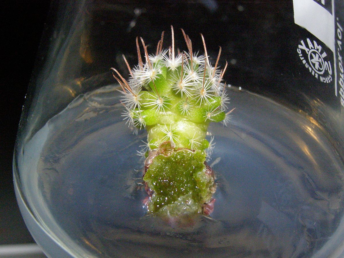
Meristem culture is a lab technique that involves culturing plant tissues to produce disease-free plants and propagate them quickly. It's a modern method of plant cloning that uses actively dividing plant tissues called meristems.


Here are some steps in the meristem culture process:
  1. Excise the meristem: Remove a small piece of the growing tip of a plant's shoot or root, which contains meristematic cells.
  2. Sterilize: Surface sterilize the excised piece.
  3. Culture: Place the meristem in a liquid or solid culture medium.
  4. Regenerate: Grow the meristem into a plantlet, then divide it and add plant hormones to regenerate more plants.
Meristem culture has many benefits, including:
  • Eliminating viruses and parasites: Meristems are the only plant tissues that can be free of viruses from an infected plant.
  • Preserving germplasm: Meristem culture can preserve germplasm characters.
1731980174446.png


Im going to thoroughly explore this topic and hopefully share some success stories with you!
Hope you enjoy the ride and find some usefulness in this thread as well :huggg:
 
Last edited:

acespicoli

Well-known member

References​

  1. Nehra N.S., Kartha K.K. (1994) Meristem and Shoot Tip Culture: Requirements and Applications. In: Vasil I.K., Thorpe T.A. (eds) Plant Cell and Tissue Culture. Springer, Dordrecht. https://doi.org/10.1007/978-94-017-2681-8_3.
  2. Varveri, C., Maliogka, V. I., & Kapari-Isaia, T. (2015). Principles for Supplying Virus-Tested Material. Control of Plant Virus Diseases - Vegetatively-Propagated Crops, 1–32. doi:10.1016/bs.aivir.2014.10.004.
  3. Bhatia, S., & Sharma, K. (2015). Micropropagation. Modern Applications of Plant Biotechnology in Pharmaceutical Sciences, 361–368. doi:10.1016/b978-0-12-802221-4.00011-x.
  4. Spangenberg, G., Wang, Z.-Y., & Potrykus, I. (1998). Meristem Culture. Biotechnology in Forage and Turf Grass Improvement, 7–17. doi:10.1007/978-3-642-72051-2_2
  5. https://byjus.com/biology/meristematic-tissue/
  6. https://www.slideshare.net/sumanrawte/meristem-cul...
📚
 

acespicoli

Well-known member
This review highlights the recent advances in plant stem cell culture and understands the cambial cells induction and culture mechanisms.


7.1 MERISTEM MORPHOLOGY​


Learning objectives
By the end of this lesson you will be able to:
  • Differentiate between primary growth from apical meristems and secondary growth from lateral meristems.
  • Describe two types of lateral meristems and the types of tissues that are derived from these meristems.

Primary growth from meristems​

You’ll recall that the apical meristem is the site of cell division and new cell production at the tips of the plant stems and roots. The cells that make up the meristem are undergoing mitotic cell division to produce more cells. These new cells result in growth and development of plant tissues. (If you haven’t previously studied mitosis, you’ll have the opportunity to do so during this class.) For now it is sufficient to know that mitosis is the process of cell division where one plant cell divides into two identical cells.


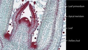
Microscope view - coleus shoot tip with labels Microscope view of coleus shoot tip with labels. BlueRidgeKitties, CC BY-NC-SA 2.0
Above is a micrograph of a coleus shoot tip. You can see the dome of the apical meristem at the very tip of the shoot surrounded by leaf primordia (rudimentary leaves). On the far left and far right are the cells of two growing leaves. You can see a trace of vascular tissue on the left leaf near the “L” of the leaf label. There is another red stained area called the axillary bud, which we’ve studied previously. The axillary bud is another very small shoot tip with a meristematic area. Axillary buds are found at a node and typically occur where a leaf petiole attaches to a stem. The axillary buds in this stage of growth are inactive, but in time may begin active cell division and develop into new branches off of the main stem.

The coleus micrograph is clearly stem tissue because you can see leaves and leaf primordia, so where are the nodes and internodes? The region where the leaves are attached, and where you find the axillary buds, is a node. Above this is the internode, and at the top where you find the leaf primordia is another node.


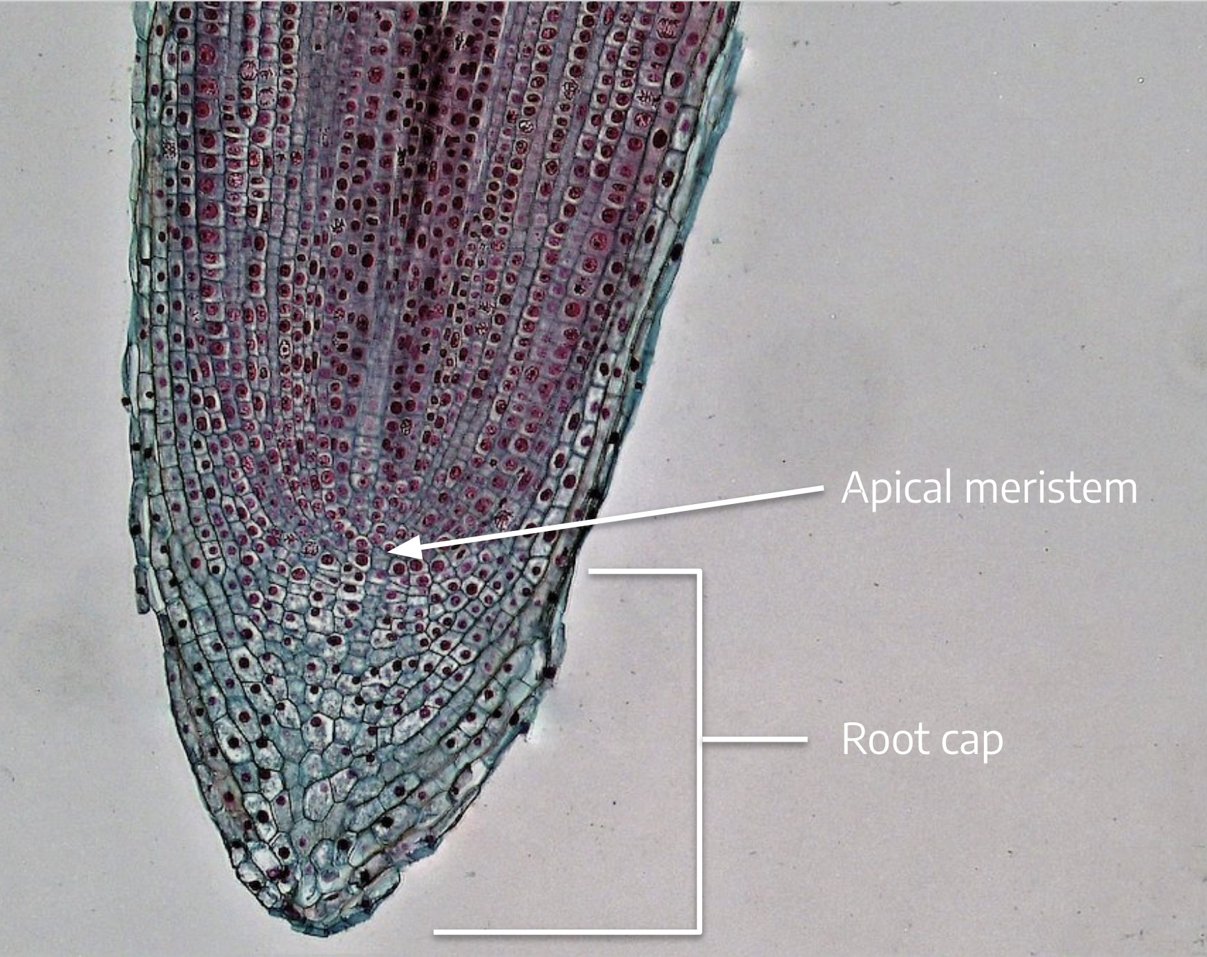
Microscope view - root tip with labels Apical meristem with root cap. Berkshire Community College, Public Domain.
The root meristem looks very different from the shoot apical meristem. Recall that, unlike branches that develop at nodes, lateral roots are formed adventitiously, as the result of meristematic activity in the pericycle cells of the root’s vascular system in the zone of maturation. We don’t see a node-internode structure like we saw with the coleus shoot tip.

When meristem cells divide, whether in the shoot or the root, one of the two resulting sister cells typically continues to be a meristem cell. The other sister cell divides a few more times and then differentiates into dermal, cortex, or vascular tissue in the stem or root. Meristem cells that remain meristematic are called initials because they continue to divide, producing new cells. The other sister cells that divide once or twice more and then differentiate are called derivative cells. The xylem and phloem tissues that result from differentiation of derivative cells are called primary xylem and primary phloem, where the word “primary” signals that the cells originated from cell divisions of the apical meristem.

To reiterate, young stems and roots have primary xylem and primary phloem that formed as a result of differentiation of derivative cells. Primary xylem and primary phloem cells trace back to an apical meristem.

Earlier you learned the arrangement of the vascular tissues in monocot and dicot stems and roots. Remember that mitotic cell divisions in the apical meristem result in lengthening of the root or shoot through production of new cells plus the elongation of those cells. With a few exceptions, this is the only type of growth — growth that is initiated by cell division in the apical meristem — you’ll find in monocots. Dicots, however, have another type of growth — from a different type of meristem — that results in thickening of the stem.


Review questions
  1. If shown a micrograph of an apical meristem, how would you determine whether it is from a root or a shoot?
  2. What happens to the initial cell mentioned in the question above? Does it continue to divide?


Secondary growth (thickening): Introducing lateral meristems​

Watch this video for a closer look at apical and lateral meristems (2:26)


Unlike annual herbaceous plants that only survive for one growing season, and whose stem and root cells trace back to cell divisions of the apical meristems, woody plants and shrubs are perennial dicots that have the capacity for secondary growth and can survive from year to year.

Some annual herbaceous dicot plants, like tomatoes, can have secondary growth, but for now let’s consider those the exceptions and focus on perennial dicot woody plants. Secondary growth is the result of activity by a special type of meristem called a lateral meristem. As with apical meristems, lateral meristems are made up of cells that undergo mitotic cell division. Mitosis in lateral meristems results in lateral growth (thickening of the stem or root) and adds to the girth of a plant rather than its length. Remember that length is the outcome of cell division in the apical meristem plus elongation of those cells. Girth or thickening is the result of lateral meristems. We’ll learn about two types of lateral meristems: vascular cambium, and cork cambium.

Vascular cambium​

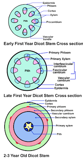
Early year dicot stem cross section and late first year dicot stem cross section. Images by Dr. Thomas L. Rost, emeritus professor at UC-Davis.
Let’s start with the vascular cambium.

The three drawings on the right show a cross section of a stem for an imaginary woody dicot. The top drawing illustrates the stem early in the first year of growth, and shows the vascular cylinders arranged in a ring around the stem. The phloem is oriented to the outside, the xylem to the inside. A thin layer of parenchyma cells between the xylem and phloem has differentiated into the fascicular cambium (fascicular refers to bundles, in this case, cambium in the vascular bundles). The fascicular cambium is meristematic and can divide to produce new phloem toward the outside and new xylem to the inside. The new xylem and phloem produced by the cambium are called 2o (secondary) xylem and 2o phloem. Recall that the original xylem and phloem that differentiated from the apical meristem’s derivative cells are called the 1o (primary) xylem and 1o phloem.

The middle drawing is of the same stem later in the year. The cortex (cortical) parenchyma cells that lay between the vascular cylinders directly in line with the fascicular cambium begin to differentiate into a type of cambium called interfascicular cambium (cambium between the bundles). This is symbolized by the line connecting the vascular cylinders. This cambium is also meristematic, and produces 2o xylem and 2o phloem.

The cross section on the bottom illustrates the stem in its second or third year of growth, when there is a noticeable buildup of 2o xylem and 2o phloem with remnants of 1o xylem and 1o phloem.

In summary, the vascular cambium is a lateral meristem formed by differentiation of parenchyma cells located between the primary xylem and phloem into fascicular cambium, followed by differentiation of cortical parenchyma between the vascular cylinders into interfascicular cambium. After a few years of secondary growth, fascicular and interfascicular cambium can no longer be distinguished, and it is all simply known as vascular cambium. This layer of cambium runs vertically (assuming that the stem is oriented vertically) and parallel to the surface of the woody stem.

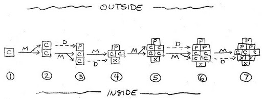
The illustration below shows how the cambium divides to produce 2o xylem and 2o phloem, with the outside of the stem toward the top of the page. Frame #1 shows a single cambium cell (C). This cell divides mitotically (M) to form two cambium daughter cells (Frame #2). Frame #3 shows that the cambium cell on the top differentiates (D) into a phloem cell (P-toward the outside) and the other cambium cell divides mitotically (M).

Cell division and differentiation from cambium to xylem/phloem Cell division and differentiation from cambium to xylem/phloem. Tom Michaels
This type of cell division, in which new cells are formed either to the outside or inside, and the cell wall that separates the two new cells is parallel to the outside of the stem, is called periclinal division. Periclinal division by the cambium makes new cells that add girth to the plant. The cells that are added subsequently differentiate into xylem and phloem depending on their location to the outside or inside of the cambium. The meristem needs to divide periclinally to add girth to the plant stem.

In Frame #4, pay particular attention to a different type of cell division, where the cambium cell has divided so that the wall between the two cells is perpendicular to the outside of the stem. This is called anticlinal division. The meristem occasionally needs to divide anticlinally because as the stem is growing in girth, the diameter of the ring of vascular cambium must expand to keep up, or it will split into pieces and no longer form a continuous ring around the stem. Frame #4 also shows that the cambium cell to the inside has differentiated into xylem (X). In Frame #5, the two cambial cells that formed from anticlinal division now each divide periclinally.
 

Latest posts

Latest posts

Top