What's new
  • ICMag with help from Landrace Warden and The Vault is running a NEW contest in November! You can check it here. Prizes are seeds & forum premium access. Come join in!

Olfactory Art

Is there value added in strains with pleasing scent qualities, to this end wish to ask more

  • All I care about is THC at the expense of terpenes (highest thc dont care how it smells or tastes )

    Votes: 0 0.0%
  • I want a THC strain that smells and tastes great

    Votes: 9 75.0%
  • I make dry sift so I want just pure THC no other stuff

    Votes: 0 0.0%
  • I smoke flowers terpenes make it taste/smell so much better

    Votes: 8 66.7%
  • I make guarda or charas so sticky/oily or waxy is better

    Votes: 1 8.3%
  • wanna see another choice added? Request it in a post or DM me

    Votes: 0 0.0%

  • Total voters
    12

IC GLASS

Member
Reasearhing terpene chemical compounds the oxygen hydrogen and carbon elements are consistant with all terpenes, the skunky and garlic, onion type all have S bond sulfur bond so it had to be present for that to manifest as a thiol or mercaptan or allicin type odor.
Gypsum is a additive I see suggested for helping with powery mildew and sulfur is a fungicide. So yeah it all makes great sense keen observation on your part. :huggg:
That’s a really insightful observation! It’s fascinating how those sulfur bonds can create such distinct odors, like skunky or garlicky smells. Plus, using gypsum and sulfur as additives for things like powdery mildew makes a lot of sense in that context—nature has some clever ways to balance things out!
 

acespicoli

Well-known member
1730259344895.png

Table of Contents

  1. Four Key Rules for Aromaticity
  2. Condition #1 for Aromaticity: The Molecule Must Be Cyclic
  3. Condition #2: Every atom in the ring must be conjugated
  4. Condition #3: The Molecule Must Have [4n+2] Pi Electrons
  5. Which Electrons Count As “Pi Electrons”?
  6. Pyridine and the Benzene Anion
  7. Some Examples With 5-Membered Rings
  8. Condition #4: The Molecule Must Be Flat
  9. Summary: Rules For Aromaticity
  10. Notes
  11. (Advanced) References and Further Reading
 

acespicoli

Well-known member
Olfactory receptor neurons (ORNs) are bipolar cells that detect and transmit odor information to the brain:


  • Location
    ORNs are found in the olfactory epithelium, a sheet of neurons and supporting cells that lines the upper nasal cavity, nasal septum, and superior nasal concha.


  • Structure
    ORNs have a small, unmyelinated axon and cilia that extend from the neuron's surface.


    • Function
      When airborne molecules bind to the cilia, the ORNs are activated and transmit information to the brain.

    • How they work
      ORNs express receptors and signaling molecules that allow them to form specific connections within the olfactory bulb. When an odorant binds to a receptor molecule, it activates a G protein, which causes a chain of events that leads to the generation of action potentials.

    • Characteristics
      ORNs exhibit different sensitivities to different odorants, and can be activated by multiple odorants or by a single odorant at different concentrations.

      1730259576111.png
    • 1730259712103.png

    • 1730259849004.png
1730260151907.png

The organization of the main olfactory system has been regarded to as a labeled line. Sensory neurons expressing the same receptor converge their afferences into single glomeruli, where synaptic contact is established with only one M/T cell, in a one-to-one topology. Thus, each olfactory receptor is represented by a single M/T cell. The accessory olfactory system, however, is integrative in the sense that each sensory neuron sends projections to several glomeruli and each M/T cell contacts several glomeruli. Although M/T cells contact glomeruli receiving afferences from different receptors, they integrate glomeruli from the same V1R or V2R subpopulation, most probably from closely related receptors. This dichotomic segregation suggests that no M/T cell contact glomeruli from both subpopulations. The non-exclusive segregation of vomeronasal afferences of the tenrec is characterized by the integration of afferences from both subpopulations into single glomeruli, suggesting that AOB neurons (such as M/T or periglomerular) may also make synaptic contact with both subpopulations.​


Number of distinguishable odors​

A widely publicized study suggested that humans can detect more than one trillion different odors.[13]

Humans can discriminate more than 1 trillion olfactory stimuli​

C Bushdid 1, M O Magnasco, L B Vosshall, A Keller
Affiliations expand
1730260735010.png

https://pubmed.ncbi.nlm.nih.gov/24653035/
 
Last edited:

bigtacofarmer

Well-known member
Veteran
I want a variety with a complex delicious flavor that dances around my senses and keeps me coming back for more. If this variety does not have a highly desirable long lasting high it will quickly become pointless to me other than maybe to breed with a variety with a great and long lasting high. I tend to not consider feeling lethargic, tired and unmotivated as being high.
 

acespicoli

Well-known member
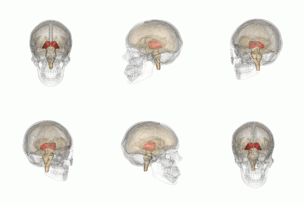
Thalamus_small.gif
1730260940717.png

1730261168443.png


The thalamus (pl.: thalami; from Greek θάλαμος, "chamber") is a large mass of gray matter on the lateral walls of the third ventricle forming the dorsal part of the diencephalon (a division of the forebrain). Nerve fibers project out of the thalamus to the cerebral cortex in all directions,

1730261347146.png

known as the thalamocortical radiations, allowing hub-like exchanges of information. It has several functions, such as the relaying of sensory and motor signals to the cerebral cortex[1][2] and the regulation of consciousness, sleep, and alertness.[3][4]
1730261594894.png

It is proposed that core relay cells and matrix cells projecting from the dorsal thalamus allow for synchronization of cortical and thalamic cells during "high-frequency oscillations that underlie discrete conscious events",[7] though this is a heavily debated area of research.
1730261678770.png
 
Last edited:

acespicoli

Well-known member
I want a variety with a complex delicious flavor that dances around my senses and keeps me coming back for more. If this variety does not have a highly desirable long lasting high it will quickly become pointless to me other than maybe to breed with a variety with a great and long lasting high. I tend to not consider feeling lethargic, tired and unmotivated as being high.
Interestingly brilliant,
it becomes clear at this moment what it is that gives strains different effects
 

I Care

Well-known member
I would best describe the desired smells for me as not to be on the nose, but in the throat and in the lung.

Like citrusy and some fruity smells seem like they’re right on the nose and The more medically viable smells make it right past the tip of the bows undetected and I can taste them maybe more than to say I smell them.

Some smells go to my eyes, some smells go to my forehead, some smells go no further than my nose. The ones that are proving good puffage without negative effects are the ones that bypass the head when smelled and seem like they go straight to taste buds and to my shoulders before even using.

Today I realize why people have been preaching against with all of these highly smelly herbs that are out there now. They almost give me an allergic reaction because they are so smelly and it seems like that really all they have. No grease, no funk, just all vape juice is how I would like to describe it.

I’ve been smoking homegrown bubba kush, homegrown sour kush, homegrown northern lights and some other stuff people have given me in recent weeks. Then somebody gave me some heavy gelato terpy all frost and flavor and no grease buds, it’s just totally subpar as far as the high goes. Just seems more like a novelty than actually having any real beneficial contribution to my mind and body and there’s actually no diversity in the flavor profile. it’s just all color food syrup sugar candy, basically feels like I over ate a bunch of candy too when the effect sets into my brain.
 

acespicoli

Well-known member
There is this essential oil booth in the mall,
you can go there and the guy who owns the booth
can give you any one of hundreds of off the shelf name brand colognes/perfumes
you would pay top dollar for, in a knock off 3 for 10$ bottle.

Went there one time and sampled thru smelling many scents just not finding what I wanted.
He patiently watched told me if I need help with anything let him know.
After not finding anything really special and since I still had many at home.
Felt as tho I may just pass this trip on the oil purchase.
Then he mentioned a thing I didnt even know was possible...

He would mix me my own custom scented essential oil cologne based on my preferences
I imagine a strain that soothes my senses custom selected he pulls out several bulk bottle to mix
All based on my said preferences and likes, offers many other samples and fine tunes it
Now I have it my perfected own designer cologne 🤷‍♂️ why not the same in a strain?

We all know which ones we favor? But the question remains why? Further whats the terpene recipe ?


1730289524644.png

How many hundreds..., thousands? ... of years or more have plants been working for us to this end ?
Just a thought :thinking:
 

acespicoli

Well-known member
Maybe some here have already experienced custom vape or terpene recipes.
Personally I have not tried those as of yet, id be keen on hearing reading those experiences.

There was one essential oil blend I found quite intriguing and here is a recipe was able to find for it .

Black Opium Inspired Recipe​


  • 6 drops of Vanilla
  • 4 drops of Coffee
  • 3 drops of Ylang Ylang
  • 2 drops of Cedarwood
  • 2 drops of Jasmine
  • 2 drops of Patchouli
  • 1 drop of Pink Pepper
  • 1 drop of Star Anise

Other Information​


  • Aromatic Profile: Resinous 39.4%, Floral 32.4%, Herbaceous 11.3%, Spicy 8.5%, Woody 8.5%
  • Emotion: Slightly Calming—with score of 31
I find Jasmine to be a heavenly scent
1730290168134.png
 

acespicoli

Well-known member
The vomeronasal organ (VNO), or Jacobson's organ, is the paired auxiliary olfactory (smell) sense organ located in the soft tissue of the nasal septum, in the nasal cavity just above the roof of the mouth (the hard palate) in various tetrapods.[1] The name is derived from the fact that it lies adjacent to the unpaired vomer bone (from Latin vomer 'plowshare', for its shape) in the nasal septum. It is present and functional in all snakes and lizards, and in many mammals, including cats, dogs, cattle, pigs, and some primates.

Some humans may have physical remnants of a VNO, but it is vestigial and non-functional.

*​

A​

D​

E​

G​

I​

L​

M​

N​

O​

P​

S​

T​

V​


Apocrine​

Main article: Apocrine sweat gland
Apocrine sweat glands are found in the armpit, areola (around the nipples), perineum (between the anus and genitals), in the ear, and the eyelids. The secretory portion is larger than that of eccrine glands (making them larger overall). Rather than opening directly onto the surface of the skin, apocrine glands secrete sweat into the pilary canal of the hair follicle. [8]

Before puberty, the apocrine sweat glands are inactive;[32] hormonal changes in puberty cause the glands to increase in size and begin functioning.[33] The substance secreted is thicker than eccrine sweat and provides nutrients for bacteria on the skin: the bacteria's decomposition of sweat is what creates the acrid odor.[34] Apocrine sweat glands are most active in times of stress and sexual excitement.[35]

In mammals (including humans), apocrine sweat contains pheromone-like compounds to attract other organisms within their species. Study of human sweat has revealed differences between men and women in apocrine secretions and bacteria.[36]

Mammary glands use apocrine secretion to produce milk.[45]
 

acespicoli

Well-known member
Lactic acid fermentation is a metabolic process by which glucose or other six-carbon sugars (also, disaccharides of six-carbon sugars, e.g. sucrose or lactose) are converted into cellular energy and the metabolite lactate, which is lactic acid in solution. It is an anaerobic fermentation reaction that occurs in some bacteria and animal cells, such as muscle cells.[1][2][3][page needed]

If oxygen is present in the cell, many organisms will bypass fermentation and undergo cellular respiration; however, facultative anaerobic organisms will both ferment and undergo respiration in the presence of oxygen.[3] Sometimes even when oxygen is present and aerobic metabolism is happening in the mitochondria, if pyruvate is building up faster than it can be metabolized, the fermentation will happen anyway.

Lactate dehydrogenase catalyzes the interconversion of pyruvate and lactate with concomitant interconversion of NADH and NAD+.

In homolactic fermentation, one molecule of glucose is ultimately converted to two molecules of lactic acid. Heterolactic fermentation, by contrast, yields carbon dioxide and ethanol in addition to lactic acid, in a process called the phosphoketolase pathway.[1]
 

acespicoli

Well-known member
1730293176086.png


1730293100042.png

Insect sex pheromone production in yeasts and plants​

https://doi.org/10.1016/j.copbio.2020.07.011


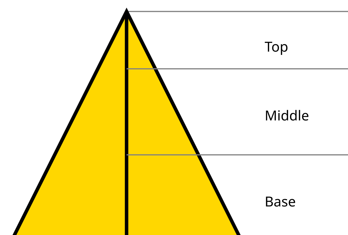
Notes in perfumery are descriptors of scents that can be sensed upon the application of a perfume. Notes are separated into three classes: top/head notes, middle/heart notes, and base/soul notes; which denote groups of scents which can be sensed with respect to the time after the application of a perfume. These notes are created with knowledge of the evaporation process and intended use of the perfume. The presence of one note may alter the perception of another—for instance, the presence of certain base or heart notes will alter the scent perceived when the top notes are strongest, and likewise the scent of base notes in the dry-down will often be altered depending on the smells of the heart notes.

The idea of notes is used primarily for the marketing of fine fragrances. The term is sometimes used by perfumers to describe approximately scents or the perfumery process to laypeople.

Volatility grouping​

Fragrant materials are listed by Poucher[1] in order of volatility and are grouped under respective evaporation coefficients (perfume notes) that range from 1 to 100.

NoteEvaporation coefficient
Top notes1 to 14 (most volatile)
Middle notes15 to 60
Base notes61 to 100 (least volatile)

Top notes​

Top notes are otherwise called the head notes.

Perceived immediately upon application of a perfume, top notes consist of small, light molecules that evaporate quickly. They form a person's initial impression of a perfume and thus are very important in the selling of the product. The scents of this note class are usually described as "fresh", "assertive" or "sharp". The compounds that contribute to top notes are strong in scent, very volatile, and evaporate quickly.

Although not as saliently perceived, the heart and base-notes contribute much to the scent in the top notes.

Citrus and ginger scents are common top notes.

Middle notes​

Also called the "heart notes", the middle notes are the scent of a perfume that emerges just before the top notes dissipate. The middle note compounds form the "heart" or main body of a perfume and emerge in the middle of the perfume's dispersion process. They serve to mask the often unpleasant initial impression of base notes, which become more pleasant with time.[citation needed] The scent of middle note compounds is usually more mellow and "rounded". Scents from this note class disappear anywhere from twenty minutes to one hour after the application of a perfume.[citation needed]

Lavender and rose scents are typical middle notes.

Base notes​

Also called the "soul notes", base notes are the scent of a perfume that appears close to the departure of the middle notes. The base and middle notes together are the main theme of a perfume. Base notes bring depth and solidity to a perfume. Compounds of this class are often the fixatives used to hold and boost the strength of the lighter top and middle notes. Consisting of large, heavy molecules that evaporate slowly, compounds of this class of scents are typically rich and "deep" and are usually not perceived until 30 minutes after the application of the perfume or during the period of perfume dry-down.

Some base notes can still be detectable in excess of twenty-four hours after application, particularly the animalic and musk notes.


acespicoli

Well-known member

Why Are We so Scent-Imental? Studying Odor-Linked Memories​

main.jpg

Abstract​

Memories that are brought on by smells are called odor-linked memories. Odor-linked memories have a large impact on our lives. When these memories are positive, our physical well-being and emotional and mental states improve. Although we often look at pictures to remember the past, odors are actually better at helping us remember. Brain scans show that odors bring on strong memories because of the brain regions that process them. The group of brain areas that are best known for processing emotions, learning, and memory also process odors. When you smell something, to process the smell, your brain uses the same areas that it would use to process emotions and memories. This makes smells great at helping us remember emotional memories! Understanding the effects of odor-linked memories can help us use them to positively affect our daily lives, in both the short-term and the long-term.
Are there particular smells that always bring you joy? When you smell certain things, such as your mother’s perfume or your favorite meal, those smells can bring up pleasant memories. These are called odor-linked memories because the memories are brought on by smells (odors). The experience that odor-linked memories cause is called the Proust effect

The experience that is brought on by memories that are odor-linked, or associated with smells
. This name came about because the author Marcel Proust wrote in his book, Swann’s Way,that the smell of a pastry he dipped in his tea brought on a rush of joy associated with his childhood. This claim made researchers curious to know whether there was a scientific explanation for Mr. Proust’s experience.

What Is the Connection Between Smells and Memory?​

Olfaction

The sense of smell. The adjective form, olfactory, means related to the sense of smell.
is just a fancy word for our sense of smell. Even though the connection between olfaction and memory is rarely thought about [1], it is actually very important. Odor-linked memories can be extremely powerful. In the laboratory, researchers ask participants to smell different odors and see what sorts of memories come to mind. The researchers then ask follow-up questions about each memory, such as whether it is clear vs. fuzzy or emotional vs. boring. It turns out that olfactory cues (smells or odors) are more effective at triggering clear and emotional memories than visual cues, such as images or photographs [2]. In fact, older adults can remember much older memories with a smell than with pictures or words [3]. So, if Mr. Proust saw a picture of his favorite pastry and the tea that he dipped it in, he might not have had such a strong experience connecting him to his childhood memories.

Researchers can use brain scans called functional magnetic resonance imaging (fMRI)

A technique for measuring brain activity as people do mental tasks. fMRI can show which areas of the brain help process different types of thoughts.
to measure brain activity while people perform mental tasks. Because fMRI looks at how the brain functions [4], it can help researchers see which parts of the brain are more (or less) involved while people remember the past in response to odors or pictures. Do odor-linked memories cause different brain activation than picture-linked memories? Yes! fMRI evidence shows that the areas of the brain that process emotions are more active when people have odor-linked memories than memories brought on by pictures (Figure 1) [5].

Figure 1 - An fMRI scan showing the difference in brain activity for odor-linked and picture-linked memories.

  • Figure 1 - An fMRI scan showing the difference in brain activity for odor-linked and picture-linked memories.
  • This image shows a scan of the brain from the top of someone’s head, as if you were looking down at them from above. The white outline shows the parts of the brain that include the amygdala and hippocampus. The colors show how big the difference in brain activity was when people had memories in response to odors and pictures, with yellow showing the biggest difference. In this study, odors caused more brain activity than pictures [Image credit: [3]].
Years of research helped us to understand that the emotional processing areas in the brain—the same areas activated by odors—include the amygdala

(sounds like “uh-mig-duh-luh”) A part of the brain involved with processing emotional memories and experiences.
and the hippocampus
A part of the brain involved with learning, especially associative learning.
, which are located in the temporal lobes
The parts of the brain that the hippocampus and amygdala are found in. This brain area is involved in processing emotions.
(Figure 2). The temporal lobes are under your skull near your left and right temples. The hippocampus is a seahorse-shaped brain area that is involved with associative learning
Learning that occurs when you connect two separate events or things together. For example, linking the smell of coffee to spending time with your grandparent.
, which is learning that occurs when you connect two separate events together. Odor-linked memory relies on associative learning because we associate (or link) the odor that we smell with the time(s) in our lives when we previously smelled it. As you can see in Figure 2, the amygdala and the hippocampus are really close together, which makes it easy for us to learn and remember emotional memories.

Figure 2 - The amygdala and hippocampus are located in the temporal lobes, with one on each side of the brain.

  • Figure 2 - The amygdala and hippocampus are located in the temporal lobes, with one on each side of the brain.
  • This image shows the left side of the brain.
How do smells get to the brain, though? After entering the nose and passing through the olfactory bulb

The part of the brain that receives information about smells.
(Figure 3), smell information is sent to the amygdala and hippocampus. Olfaction is the only sense that gets processed in this brain area. Therefore, smell is the sense most strongly linked to brain areas that are involved in emotion, learning, and memory. Thanks to fMRI scans, we now have evidence to explain why the smell of freshly cut grass takes us back to a previous summer in our memories, or why Mr. Proust so clearly remembered his childhood just from smelling a pastry. Smelling the grass or a pastry activates the brain areas responsible for emotions and memories!

Figure 3 - Smell is sent through the olfactory bulb (yellow) to the amygdala and hippocampus areas (orange).

  • Figure 3 - Smell is sent through the olfactory bulb (yellow) to the amygdala and hippocampus areas (orange).

Mental and Physical Benefits of Odor-Linked Memories​

Odor-linked memories are special because they can do more than just transport us back to a different time in our lives. They can improve our health as well. It might seem surprising that our sense of smell can have such a positive impact on our well-being, but it is true! Researchers have found that odor-linked memories can actually be more positive than memories that are brought on by any other senses [1]. Pleasant memories put us in good moods, and being in a good mood helps to reduce stress. Stress can present itself in our bodies as inflammation (swelling, redness), which is a sign doctors look for in many different diseases [1]. Therefore, when odor-linked memories reduce our stress levels, we relax, and our physical health improves.
In addition to improving our physical health, odor-linked memories can improve our mental health. Mental health benefits of these memories include boosts to self-esteem, improved social interactions, and a sense of optimism (a sunny outlook). Odor-linked memories have also been shown to inspire self-confidence and motivation, and even to help adults quit smoking cigarettes! When smokers had pleasant odor-linked memories, they said that their cigarette cravings were reduced [1].

What Else Is There to Know?​

Our lives are affected by the smells around us in big ways. We know that certain odors can actually improve our lives in the long run, by making us healthier. You can even use what you know about odors and memory to help you in school. Because we know that odor is so strongly linked to memory, if you smell the same odor when you study for and take a test, you have a better chance of remembering what you studied! For example, if you use peppermint lip balm while studying, you should put on that same lip balm before your test. The smell of peppermint can help you remember what you studied because of associative memory. Researchers proved this using the smell of chocolate [6]! Just be sure not to use the same lip balm smell for multiple classes, so your brain can remember the right class!
What we have discovered about odor-linked memories is important, but there is still so much more to learn. Because positive memories can help lower stress, they also help improve mood and can help people change unwanted behaviors, like smoking. This means that the effects of odor-linked memories could be used in therapy and counseling, to help improve people’s lives in both the short- and long-term. In conclusion, there are many effects of odor-linked memories, but we are not done studying all the ways they affect our lives just yet. It is a good thing Mr. Proust dipped that pastry into his tea…otherwise, we might not be aware of all of the positive impacts that odor-linked memories can have!


acespicoli

Well-known member
Figure 1 - An fMRI scan showing the difference in brain activity for odor-linked and picture-linked memories.

  • Figure 1 - An fMRI scan showing the difference in brain activity for odor-linked and picture-linked memories.
  • This image shows a scan of the brain from the top of someone’s head, as if you were looking down at them from above. The white outline shows the parts of the brain that include the amygdala and hippocampus. The colors show how big the difference in brain activity was when people had memories in response to odors and pictures, with yellow showing the biggest difference. In this study, odors caused more brain activity than pictures [Image credit: [3]].

If a picture is worth a 1000 words... 🤷‍♂️
Leonardo da Vinci wrote that a poet would be "overcome by sleep and hunger before [being able to] describe with words what a painter is able to [depict] in an instant."[
 

acespicoli

Well-known member

A Window Into Your Brain: How fMRI Helps Us Understand What Is Going on Inside Our Heads​


main.jpg

Abstract​

The brain—the last frontier of modern science. Despite many technological advances, we still know little about how the brain works. Luckily, the development of a technique called functional magnetic resonance imaging (fMRI) is slowly helping change this. fMRI can measure brain activity without opening the skull or exposing the brain cells to harmful radiation. By using the blood’s magnetic properties, fMRI can detect changes in blood flow related to brain activity, allowing scientists and physicians to tell which regions of the brain are more active than others. Currently, researchers use fMRI to study various aspects of brain activity in health and disease. Scientists continue to push the boundaries of the fMRI technique and combine it with other techniques to obtain an even better understanding of brain function and dysfunction.

What is fMRI and How Does It Work?​

“Do you ever look at someone and wonder, ‘what is going on inside their head?”’ This famous question posed by the movie character Joy, from the Disney Pixar movie Inside Out, is a puzzle doctors and scientists seek to solve daily with the help of brain imaging. Brain imaging allows doctors and scientists to view the interior structures of the brain without ever opening the skull. There are several brain-imaging techniques. One of them is called magnetic resonance imaging (MRI), which looks at the structure of the brain and another is functional magnetic resonance imaging (fMRI), which looks at the brain’s function.
fMRI measures brain activity by tracking changes in blood flow to the brain. fMRI is derived from structural MRI. In fact, they both use the same machine, called a magnetic resonance scanner (Figure 1). MRI technology is used to create detailed, 3D images of the internal structure of an object using magnetic fields

Field that occurs around a magnet, which exerts a force that attracts or repels other magnetic objects.
and radio waves [1]. MRI can be used to study body parts other than the brain, and even non-living objects. For example, MRI could be used by an archaeologist to take pictures of the inside of a fossil. fMRI can also be used to image body parts other than the brain. In medicine, brain MRI and fMRI are used to help recognize illnesses, plan treatments, and study the underlying causes of diseases and disorders.

Figure 1 - Components of an MR scanner [2].

  • Figure 1 - Components of an MR scanner [2].
  • MRI and fMRI scans take place within the same MRI scanner.
MR scanners work by taking pictures of the brain one thin layer at a time. The pictures are then stacked like pancakes to create a full picture of the region being imaged. How is this possible, you may ask? The human body is made up of billions of molecules, including water molecules, which can be detected by the MRI machine. Atoms in all molecules, including water molecules (H2O), contain protons [1]. Protons are like tiny magnets [1]. In the absence of a very strong magnetic field (that is, when we are outside the MR scanner), protons in our body are oriented in random directions (Figure 2A). When we lie inside the scanner, its strong magnetic field, which is usually tens of thousands of times the strength of the earth’s magnetic field, forces these protons to align with the field, although we cannot feel this at all (Figure 2B). The gradient coil (see Figure 1) helps the scanner operators determine exactly where our body is inside the scanner.
Figure 2 - Molecules in the body during an MRI scan.

  • Figure 2 - Molecules in the body during an MRI scan.
  • (A) Molecules in the body, which act like tiny magnets, are oriented in random directions in the absence of a strong magnetic field. (B) MR scanners produce a strong magnetic field (dashed red arrow) that forces the molecules in the body to align with the field. (C) RF coils transmit radio frequencies (dotted aqua arrow) throughout areas of the body being imaged. The molecules become realigned in between the directions of the magnetic fields of the RF coil and the scanner. (D) When radio frequencies are no longer transmitted, the molecules revert, or “relax,” to their original alignment with the scanner’s magnetic field, releasing energy in the form of electromagnetic waves (inset at top right). The energy that is released can be analyzed to create an image of the body part.
Then, the radio frequency (RF) coils transmit radio frequency waves

Special electromagnetic signals created by radio frequency coils to realign protons from the magnetic field of the MRI scanner’s magnet.
throughout the areas of the body that are being imaged, to realign these protons yet again, but temporarily (Figure 2C). RF coils may be part of the MRI machine for full-body scan or worn as a special headband when just imaging the brain. When the radio frequencies are no longer transmitted, the protons “relax” into their original alignment with the scanner’s magnetic field. In doing so, the protons release the energy that was tugging them in the RF coil’s direction (like releasing a slingshot), in the form of electromagnetic signals

Signals created through the vibrations of magnetic fields and electric fields.

(Figure 2D).

In the same way that millions of water droplets can create a puddle, the signals from millions of protons, when carefully anaylzed, can come together to create a detailed image of the body [1]. While MRI only takes pictures of the brain structure, fMRI shows the activity (or function) of the brain, by comparing the blood flow under different conditions.

Neurons: the Building Blocks of Our Brains​

The brain helps us understand and respond to the world around us. It enables us to interpret things we see, touch, hear, and taste, and regulates our body’s responses to the external environment. It does all this through networks of tiny cells called neurons

Nerve cells that send and receive electrical and chemical signals across long distances, especially in the brain.
, which process and transmit information between the brain and the rest of the body [3]. When the brain is faced with a task, such as remembering an idea, neurons responsible for that activity become more active than other neurons around them. They do this by creating chemical and electrical signals and transferring them from one neuron to another. This process is referred to as neural activity or brain activity.

How Does fMRI Measure Brain Activity?​

Neural activity requires energy. Like other cells in the body, neurons generate energy by using oxygen to break down sugar. When neural activity increases in a part of the brain, more energy is used. To replenish this energy, more oxygen-carrying blood is transported to that brain region. The blood transports oxygen using a molecule called hemoglobin

Iron-containing protein inside red blood cells that captures oxygen and transports it to the tissues of the body.
. Hemoglobin contains iron, which gives it magnetic properties, like a tiny iron filing. Depending on whether hemoglobin is carrying oxygen or not (that is, whether it is oxygenated or deoxygenated), it has slightly different magnetic properties. Thus, greater neural activity results in greater flow of oxygenated blood (Figure 3), so that brain regions that are more active are slightly more magnetic. This results in slightly different patterns of electromagnetic waves.

Figure 3 - Movement of oxygenated and deoxygenated hemoglobin during neural activity.

  • Figure 3 - Movement of oxygenated and deoxygenated hemoglobin during neural activity.
  • (A) When neural activity increases in region of the brain, more energy is used by that brain region, and hemoglobin becomes deoxygenated as it gives oxygen to the cells that need it. (B) To replenish this energy, more oxygenated blood is transported to that brain region.
fMRI detects brain activity by measuring the changes in both the amount of oxygen in the blood and the amount of blood flow [4, 5]. This measurement is known as blood-oxygen-level-dependent activity (BOLD activity). In other words, BOLD activity is a convenient stand-in for brain activity: fMRI measures brain activity indirectly by measuring BOLD activity. This is somewhat akin to figuring out where and when lightning happened by listening to the thunder.

“Bold” Insights Into Brain Function​

Over the last three decades or so, researchers have using BOLD MR imaging to begin to answer Joy’s question about what is going on inside people’s heads. The power of fMRI to peer into the otherwise impenetrable depths of brain function, in humans or animals, is illustrated beautifully by the work of Dr. Gregory Berns and his colleagues. They compared the BOLD activity in the brains of dogs in response to two different hand signals by their handlers: one that told the dogs they were going to get a tasty food reward, and another that told them they would not get a reward (Figure 4A).
Figure 4 - Brain activity in dogs when they are expecting a reward [6].

  • Figure 4 - Brain activity in dogs when they are expecting a reward [6].
  • (A) A side view of a dog’s head. The blue plane shows the plane in which the brain images were obtained. (B) The BOLD activity in two healthy female dogs named Callie and McKenzie was measured while they watched their handler’s hand signals for reward vs. no-reward. fMRI images are on the left, and the structural MRI images are on the right. The images are a top-down view of the brain taken at the plane shown in (A). The caudate region (CD) lights up in the fMRI images, meaning that it is activated, and the green arrow shows the location of the CD in the corresponding structural MRI images. R and L denote the right and left sides of the dog. The color bar indicates the level of activity, with red being highest and blue being lowest.
They found that a region deep in the brain, the caudate nucleus

A region of the brain which, among other things, responds to rewards.
(CD), was more active when the dogs saw the signal for the reward than when they saw the no-reward signal (Figure 4B). Previous studies in humans and other species have shown that the same brain region is activated in humans when we are expecting a reward. So, there is an answer to Joy’s question in this example: what goes on in a dog’s head is remarkably similar to what goes on in our own heads in similar situations, and can be imaged using fMRI in both cases [6]. This remarkable insight would not have been possible without being able to “read” the dogs’ brains using fMRI!
 

acespicoli

Well-known member

Mimicry​

Main article: Mimicry

Images A and B show real wasps; the others show Batesian mimics: three hoverflies and one beetle.
Bates' work on Amazonian butterflies led him to develop the first scientific account of mimicry, especially the kind of mimicry which bears his name: Batesian mimicry.[66] This is the mimicry by a palatable species of an unpalatable or noxious species (the model), gaining a selective advantage as predators avoid the model and therefore also the mimic. Mimicry is thus an anti-predator adaptation. A common example seen in temperate gardens is the hoverfly (Syrphidae), many of which—though bearing no sting—mimic the warning coloration of aculeate Hymenoptera (wasps and bees). Such mimicry does not need to be perfect to improve the survival of the palatable species.[67]

Bates, Wallace and Fritz Müller believed that Batesian and Müllerian mimicry provided evidence for the action of natural selection, a view which is now standard amongst biologists.[68][69][70]
 

acespicoli

Well-known member
Mel Frank, Manhattan, 1970

The stuff that I grew back then that got tested by the government lab at the University of Mississippi, the highest stuff was almost 12 percent THC. They said it was the strongest thing they had tested. In fact, the guy there called me and asked me if the stuff was adulterated. But now you routinely have stuff that tests at 20 percent and over, and to me, at this point, I don’t really care about that. I care about the fragrance and flavor, ok? That’s what’s going to make your highs and also just your sensory pleasure different. Once you get into the teens what difference does it make?
Afghani 1 from 1979

-Mel Frank (When we were criminals, article)
 

Latest posts

Latest posts

Top