What's new

Clones, Mother plants, How to "Clean" cuttings!

acespicoli

Well-known member
Mentor
1732242502739.png

Cannabis cell culture, transformation, and micropropagation work since 1972–2020.
Front. Plant Sci., 02 March 2021
Sec. Crop and Product Physiology
Volume 12 - 2021 | https://doi.org/10.3389/fpls.2021.627240
1732242797769.png

Clonex Clone Solution is specifically formulated to provide rooted clones and seedlings with a special blend of the highest quality minerals including Nitrogen, Phosphorus, Potassium, and Calcium plus other essential elements that all plants require for vigorous growth. Clonex Clone Solution also contains Vitamin B1 which reduces the risk of transplant shock.

Used with Clonex Rooting Gel or any other rooting agent, it encourages rapid root development while helping to minimize stress. Clonex Clone Solution is the result of extensive horticultural research and is designed to work together with Clonex Rooting Gel or other rooting agents for outstanding results.

Clonex Clone Solution contains specific micro nutrients and root enhancing agents, carefully formulated to initiate and nourish new root cells in plants. Unlike competitive products, Clonex Clone Solution is highly concentrated.
***
1732242903170.png

Clonex Gel is a high performance, water-based, rooting compound. This tenacious gel which will remain in contact around the stem, sealing the cut tissue and supplying the hormones needed to promote root cell development and vitamins to protect the delicate new root tissue. Clonex has a full spectrum of mineral nutrients and trace elements to nourish the young roots during their important formative stages. The scientific breakthrough puts Clonex Rooting Gel years ahead of manufacturers of old fashioned hormones and powders.

Since 1988 Clonex Rooting Gel has led the way in plant propagation with billions of clones successfully rooted, including virtually every species of plant known to man. The Clonex formulation has been fine-tuned to give the explosive root development and for this reason serious growers and plant nurseries depend upon it for successful and profitable plant propagation. It is completely alcohol-free.

Clonex Rooting Gel is widely recognized as an industry standard for its ability to meet the successful and consistent results required for propagation done with vegetative cuttings. It is registered with the EPA and is approved for use on all food crops, including medicinal plants, in all 50 states plus Washington DC and Puerto Rico and is the only rooting gel approved by the Colorado Department of Agriculture for propagating medicinal plants.

HOW TO USE CLONEX ROOTING GEL:

First, dip cutting in gel to desired depth. Then insert cutting into rooting medium. Mist cuttings and place in propagator or a warm, clean, moist and humid environment. Finally, look for root development in 1 – 2 weeks.

As a matter of fact, you can use Clonex Rooting Gel on all types of plant cuttings, including woody, herbaceous, and flowering ornamental species, vegetables, fruit trees and small fruits.
1732243215566.png

1732243306801.png


1732242812111.png

CLONE KING

 
Last edited:
acespicoli

Well-known member
Mentor

Improving results​


A plastic cold frame with semi-white plastic, used to keep the cuttings humid and semi-shaded
There are ways of improving the growth of stem cutting propagations. Intensifying light allows cuttings to root and sprout faster, though the heat thus generated could cause the propagation material distress.[29] Azalea cuttings can be mildly heated in water to disinfect it from the fungus pathogen Rhizoctonia, and this could potentially be used for other plants.[30]

Soil​

Depending on the type of cutting (i.e. tree, shrub, succulent, cacti) different potting soil mixes can be used. Many commercial companies sell medium specifically for growing cuttings.

Air and soil humidity​

Although several options can be used here, usually plastic is used to cover the softwood and semi-hardwood cuttings. The soil below the trays (to increase air moisture) and the soil in the trays themselves is kept moist but not waterlogged (=completely saturated). The trays the cuttings sit in are best placed on stones to prevent capillary action (as this can keep the soil inside the trays too wet). Soil in the trays should be kept at 85 to 95% saturation.[31] Automated (overhead) misting systems, boom systems or fog systems[32] can be used in greenhouses.

A typical misting frequency during sticking and callusing includes misting for 5–8 seconds every 5–10 minutes over a 24-hour period. After 3 to 4 days, misting is reduced to 3–5 seconds every 10–20 minutes during the day and less frequently at night. When roots become visible (stage 3) misting can be reduced, and by stage 4 (toning), little to no misting should be done (by day 10 to 14 for most species[31]).[32] When using plastic tents, far less misting is needed (once or twice a day).[33] The greenhouse or cold frame should be ventilated once in a while[34] to prevent formation of molds (manually).

Air and soil temperature​

Air temperature for softwood and semi-hardwood cuttings is optimal at around 70 °F[35][36][37][38] (21.1 °C) but temperatures as low as 55 °F (12.7 °C) are acceptable. Heating the air above 75 °F (23.8 °C) stimulates the growth of pathogens.[35] Ventilating (manually or through automatic window openers) the greenhouse or cold frame can lower the air temperature. Automated thermostat systems can also be used in greenhouses to keep the heat at a specific temperature. Bottom heating (soil) tends to be ideal for root initiation since growing media temperature is best maintained at 20-22 °C.[31]

Sunlight​

Whereas cuttings need to be kept warm and some amount of light needs to be provided, it needs to be kept out of direct sunlight.[39] Some ways to accomplish this include using white wash, semi-white plastic, retractible shade curtains (which can be deployed if the sun temporarily pierces through), ... Optimum light levels are around 120 to 200 μmol/m2s at the first stage (sticking[32]). Once callus has been formed (stage 2: callusing[32]) and roots start to form and take up water (stage 3: root development phase), light intensity levels can be gradually increased (to 200 to 800 μmol/m2s).[40][41] Most propagators find that 5 to 10 moles per day (i.e. observed using a Daily Light Integral sensor) will result in a consistent rooting and growth[31]
 
Last edited:

H e d g e

Well-known member
I’ve been preparing the plant before taking cuts, pinching the stem at the node I plan to cut through and allowing a callus to form as well as trimming any lower leaves I plan on removing and letting the wounds dry out while it’s still on the plant before taking the cut.

I get pretty good results whether I do this or not though, probably not much in it either way, I just don’t like putting open wounds in high humidity as they can sometimes become infected with mould and it increases the surface area for up-taking whatever’s in the clone x making the cut through a callus.

I also put some crushed herm seeds in the soil to inoculate the roots with biology as they grow, probably over kill but it works.
 

acespicoli

Well-known member
Mentor
1732246370323.png

Effects of auxin​

Main article: auxin
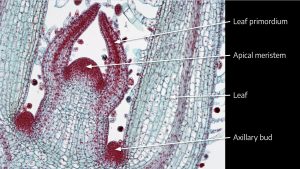
As the apical meristem grows and forms leaves, a region of meristematic cells is left behind at the node between the stem and the leaf. These axillary buds are usually dormant, inhibited by auxin produced by the apical meristem, which is known as apical dominance.

If the apical meristem is removed, or has grown a sufficient distance away from an axillary bud, the axillary bud may become activated (or more appropriately freed from hormone inhibition). Like the apical meristem, axillary buds can develop into a stem or flower.


THIS IS THE MOST SUCCESSFUL CUT IN THE WORST CONDITIONS
BROKEN BRANCH FROM MAIN STEM
 

acespicoli

Well-known member
Mentor

Apex removal​

Plant physiologists have identified four different stages the plant goes through after the apex is removed (Stages I-IV). The four stages are referred to as

  1. lateral bud formation,
  2. "imposition of inhibition" (apical dominance),
  3. initiation of lateral bud outgrowth following decapitation, and
  4. elongation and development of the lateral bud into a branch.
These stages can also be defined by the hormones that are regulating the process which are as follows:

Stage I, cytokinin promoted, causing the lateral bud to form since cytokinin plays a role in cell division;
Stage II, auxin is promoted, resulting in apical dominance ("imposition of inhibition");
Stage III, cytokinin released resulting in outward growth of the lateral bud; and
Stage IV, auxin is decreased and gibberellic acid is promoted which results in cell division, enabling the bud or branch to continue outward growth.
[1]

More simply stated, lateral bud formation is inhibited by the shoot apical meristem (SAM). The lateral bud primordium (from which the lateral bud develops) is located below SAM. The shoot tip rising from the SAM inhibits the growth of the lateral bud by repressing auxin. When the shoot is cut off, the lateral bud begins to lengthen which is mediated by a release of cytokinin. Once the apical dominance has been lifted from the plant, elongation and lateral growth is promoted and the lateral buds grow into new branches. When lateral bud formation prevents the plant from growing upward, it is undergoing lateral dominance. Often, lateral dominance can be triggered by decapitating the SAM or artificially decreasing the concentration of auxin in plant tissues.
 

acespicoli

Well-known member
Mentor
I’ve been preparing the plant before taking cuts, pinching the stem at the node I plan to cut through and allowing a callus to form as well as trimming any lower leaves I plan on removing and letting the wounds dry out while it’s still on the plant before taking the cut.

I get pretty good results whether I do this or not though, probably not much in it either way, I just don’t like putting open wounds in high humidity as they can sometimes become infected with mould and it increases the surface area for up-taking whatever’s in the clone x making the cut through a callus.

I also put some crushed herm seeds in the soil to inoculate the roots with biology as they grow, probably over kill but it works.
:yeahthats

As the apical meristem grows and forms leaves,
a region of meristematic cells is left behind at the node between the stem and the leaf.

Meristematic cells are undifferentiated plant cells that divide and replenish themselves to produce new cells that give rise to plant organs:


  • Characteristics
    Meristematic cells are small, nearly spherical, and tightly packed with thin cell walls, dense cytoplasm, and small vacuoles. They are similar to stem cells in animals.


  • Functions
    Meristematic cells play essential roles in plant physiology, including:
    • Plant growth


    • Regeneration


      • Flowering

      • Leaf and branch sprouting

      • Wound healing
    • Types
      There are three types of meristematic tissues:
        • Apical: Located at the growing shoot and root tips

        • Intercalary or basal: Located in the middle

        • Lateral: Located at the sides, also known as cambium
    • Cultivation
      Meristem cell cultivation has many applications, including:
        • Reproducing different cultivars

        • Genetically modifying plant species

        • Preserving extinct species

        • Producing virus-free plants

        • Assessing plant metabolism and physiology
 

acespicoli

Well-known member
Mentor

Anatomy​

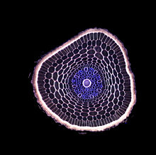
The cross-section of a barley root
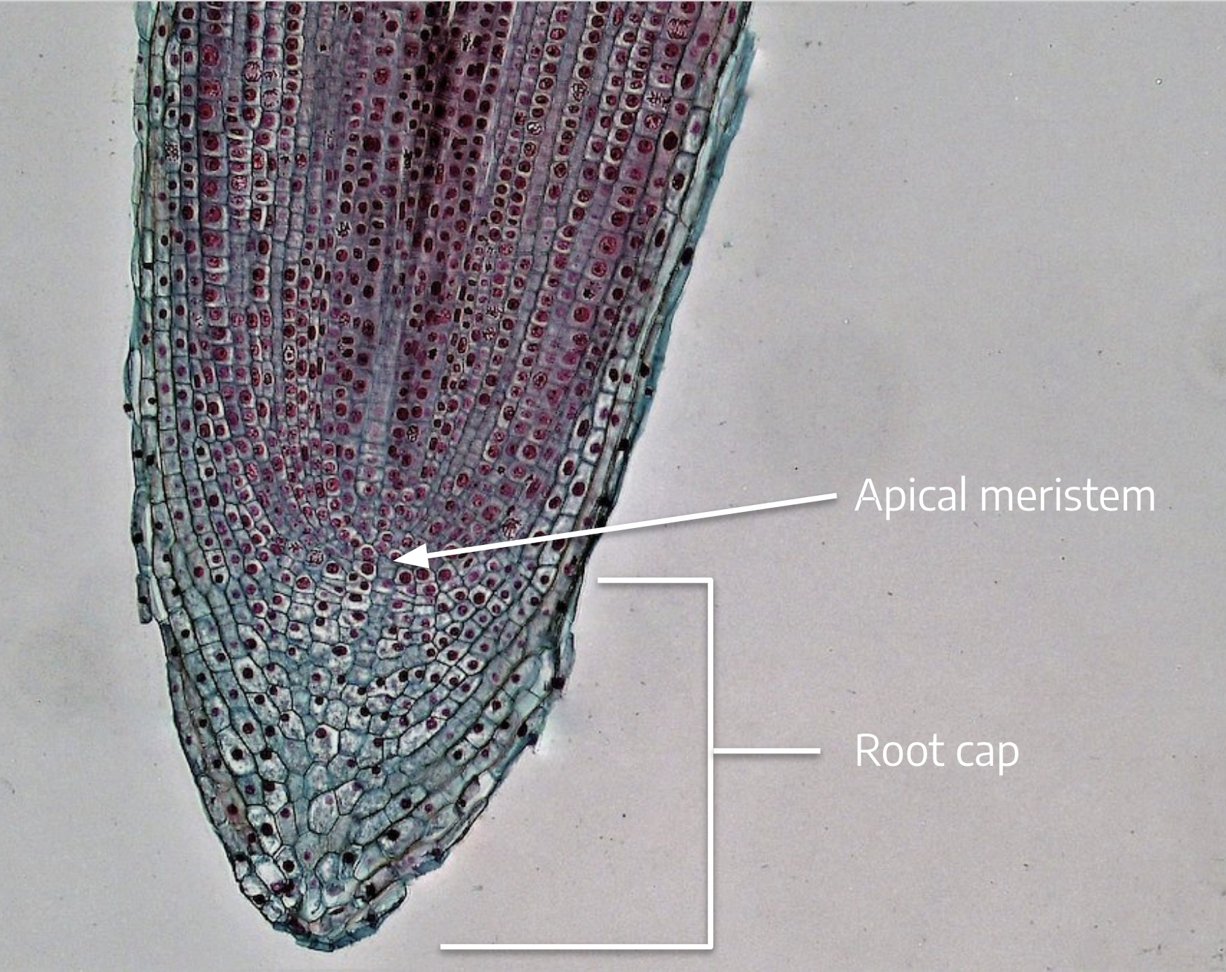
Root morphology is divided into four zones: the root cap, the apical meristem, the elongation zone, and the hair.[4] The root cap of new roots helps the root penetrate the soil. These root caps are sloughed off as the root goes deeper creating a slimy surface that provides lubrication. The apical meristem behind the root cap produces new root cells that elongate. Then, root hairs form that absorb water and mineral nutrients from the soil.[5] The first root in seed producing plants is the radicle, which expands from the plant embryo after seed germination.

When dissected, the arrangement of the cells in a root is root hair, epidermis, epiblem, cortex, endodermis, pericycle and, lastly, the vascular tissue in the centre of a root to transport the water absorbed by the root to other places of the plant.[clarification needed]

Ranunculus root cross section
Perhaps the most striking characteristic of roots that distinguishes them from other plant organs such as stem-branches and leaves is that roots have an endogenous[6] origin, i.e., they originate and develop from an inner layer of the mother axis, such as pericycle.[7] In contrast, stem-branches and leaves are exogenous, i.e., they start to develop from the cortex, an outer layer.

In response to the concentration of nutrients, roots also synthesise cytokinin, which acts as a signal as to how fast the shoots can grow. Roots often function in storage of food and nutrients. The roots of most vascular plant species enter into symbiosis with certain fungi to form mycorrhizae, and a large range of other organisms including bacteria also closely associate with roots.[8]

All components of the root architecture are regulated through a complex interaction between genetic responses and responses due to environmental stimuli. These developmental stimuli are categorised as intrinsic, the genetic and nutritional influences, or extrinsic, the environmental influences and are interpreted by signal transduction pathways.[12]

Extrinsic factors affecting root architecture include gravity, light exposure, water and oxygen, as well as the availability or lack of nitrogen, phosphorus, sulphur, aluminium and sodium chloride. The main hormones (intrinsic stimuli) and respective pathways responsible for root architecture development include:

AuxinLateral root formation, maintenance of apical dominance and adventitious root formation.
CytokininsCytokinins regulate root apical meristem size and promote lateral root elongation.
EthylenePromotes crown root formation.
GibberellinsTogether with ethylene, they promote crown primordia growth and elongation. Together with auxin, they promote root elongation. Gibberellins also inhibit lateral root primordia initiation.
 

acespicoli

Well-known member
Mentor
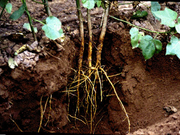
  • Adventitious roots arise out-of-sequence from the more usual root formation of branches of a primary root, and instead originate from the stem, branches, leaves, or old woody roots. They commonly occur in monocots and pteridophytes, but also in many dicots, such as clover (Trifolium), ivy (Hedera), strawberry (Fragaria) and willow (Salix). Most aerial roots and stilt roots are adventitious. In some conifers adventitious roots can form the largest part of the root system. Adventitious root formation is enhanced in many plant species during (partial) submergence, to increase gas exchange and storage of gases like oxygen.[28] Distinct types of adventitious roots can be classified and are dependent on morphology, growth dynamics and function.[29][30]
 

acespicoli

Well-known member
Mentor

Synthetic auxins​

In the course of research on auxin biology, many compounds with noticeable auxin activity were synthesized. Many of them had been found to have economical potential for human-controlled growth and development of plants in agronomy.

Auxins are toxic to plants in large concentrations; they are most toxic to dicots and less so to monocots.[38] Because of this property, synthetic auxin herbicides, including 2,4-dichlorophenoxyacetic acid (2,4-D) and 2,4,5-trichlorophenoxyacetic acid (2,4,5-T), have been developed and used for weed control.

However, some exogenously synthesized auxins, especially 1-naphthaleneacetic acid (NAA) and indole-3-butyric acid (IBA), are also commonly applied to stimulate root growth when taking cuttings of plants or for different agricultural purposes such as the prevention of fruit drop in orchards.

Used in high doses, auxin stimulates the production of ethylene, also a native plant hormone. Excess ethylene can inhibit elongation growth, cause leaves to fall (abscission), and even kill the plant. Some synthetic auxins, such as 2,4-D and 2,4,5-T are marketed also as herbicides. Dicots, such as dandelions, are much more susceptible to auxins than monocots, such as grasses and cereal crops. So these synthetic auxins are valuable as synthetic herbicides. 2,4-D was the first widely used herbicide, and it is still in use.[39] 2,4-D was first commercialized by the Sherwin-Williams company and saw use in the late 1940s. It is easy and inexpensive to manufacture.


Root growth and development​

Auxins promote root initiation.[32] Auxin induces both growth of pre-existing roots and root branching (lateral root initiation), and also adventitious root formation. As more native auxin is transported down the stem to the roots, the overall development of the roots is stimulated. If the source of auxin is removed, such as by trimming the tips of stems, the roots are less stimulated accordingly, and growth of stem is supported instead.

In horticulture, auxins, especially NAA and IBA, are commonly applied to stimulate root initiation when rooting cuttings of plants. However, high concentrations of auxin inhibit root elongation and instead enhance adventitious root formation. Removal of the root tip can lead to inhibition of secondary root formation.

Apical dominance​

Main article: Apical dominance
Auxin induces shoot apical dominance; the axillary buds are inhibited by auxin, as a high concentration of auxin directly stimulates ethylene synthesis in axillary buds, causing inhibition of their growth and potentiation of apical dominance. When the apex of the plant is removed, the inhibitory effect is removed and the growth of lateral buds is enhanced. This is called decapitation, usually performed in tea plantations and hedge-making. Auxin is sent to the part of the plant facing away from the light, where it promotes cell elongation, thus causing the plant to bend towards the light.[33]
 

acespicoli

Well-known member
Mentor
I’ve been preparing the plant before taking cuts, pinching the stem at the node I plan to cut through and allowing a callus to form as well as trimming any lower leaves I plan on removing and letting the wounds dry out while it’s still on the plant before taking the cut.
:yeahthats

screenshot-drive_google_com-2024_11_21-22_42_57.png

Truly a superior method macerated node cuts
poor example of rooting but a accurate demo of technique


The vascular cambium is the main growth tissue in the stems and roots of many plants,
It produces secondary xylem inwards, towards the pith, and secondary phloem outwards,
towards the bark.
1732249131883.png

The cells of the vascular cambium (F) divide to form phloem on the outside, located beneath the bundle cap (E), and xylem (D) on the inside. Most of the vascular cambium is here in vascular bundles (ovals of phloem and xylem together) but it is starting to join these up as at point F between the bundles.


keyword = meristematic
Differentiated plant cells generally cannot divide or produce cells of a different type. Meristematic cells are undifferentiated or incompletely differentiated. They are totipotent and capable of continued cell division. Division of meristematic cells provides new cells for expansion and differentiation of tissues and the initiation of new organs, providing the basic structure of the plant body.


In dicots, layer two of the corpus determines the characteristics of the edge of the leaf. The corpus and tunica play a critical part of the plant physical appearance as all plant cells are formed from the meristems. Apical meristems are found in two locations: the root and the stem.

Apical dominance is where one meristem prevents or inhibits the growth of other meristems.


Propagating through cuttings is another form of vegetative propagation that initiates root or shoot production from secondary meristematic cambial cells. This explains why basal 'wounding' of shoot-borne cuttings often aids root formation.[32]
 
Last edited:

acespicoli

Well-known member
Mentor

7.1 MERISTEM MORPHOLOGY​


Learning objectives
By the end of this lesson you will be able to:
  • Differentiate between primary growth from apical meristems and secondary growth from lateral meristems.
  • Describe two types of lateral meristems and the types of tissues that are derived from these meristems.

Primary growth from meristems​

You’ll recall that the apical meristem is the site of cell division and new cell production at the tips of the plant stems and roots. The cells that make up the meristem are undergoing mitotic cell division to produce more cells. These new cells result in growth and development of plant tissues. (If you haven’t previously studied mitosis, you’ll have the opportunity to do so during this class.) For now it is sufficient to know that mitosis is the process of cell division where one plant cell divides into two identical cells.


Microscope view - coleus shoot tip with labels Microscope view of coleus shoot tip with labels. BlueRidgeKitties, CC BY-NC-SA 2.0
Above is a micrograph of a coleus shoot tip. You can see the dome of the apical meristem at the very tip of the shoot surrounded by leaf primordia (rudimentary leaves). On the far left and far right are the cells of two growing leaves. You can see a trace of vascular tissue on the left leaf near the “L” of the leaf label. There is another red stained area called the axillary bud, which we’ve studied previously. The axillary bud is another very small shoot tip with a meristematic area. Axillary buds are found at a node and typically occur where a leaf petiole attaches to a stem. The axillary buds in this stage of growth are inactive, but in time may begin active cell division and develop into new branches off of the main stem.

The coleus micrograph is clearly stem tissue because you can see leaves and leaf primordia, so where are the nodes and internodes? The region where the leaves are attached, and where you find the axillary buds, is a node. Above this is the internode, and at the top where you find the leaf primordia is another node.


Microscope view - root tip with labels Apical meristem with root cap. Berkshire Community College, Public Domain.
The root meristem looks very different from the shoot apical meristem. Recall that, unlike branches that develop at nodes, lateral roots are formed adventitiously, as the result of meristematic activity in the pericycle cells of the root’s vascular system in the zone of maturation. We don’t see a node-internode structure like we saw with the coleus shoot tip.

When meristem cells divide, whether in the shoot or the root, one of the two resulting sister cells typically continues to be a meristem cell. The other sister cell divides a few more times and then differentiates into dermal, cortex, or vascular tissue in the stem or root. Meristem cells that remain meristematic are called initials because they continue to divide, producing new cells. The other sister cells that divide once or twice more and then differentiate are called derivative cells. The xylem and phloem tissues that result from differentiation of derivative cells are called primary xylem and primary phloem, where the word “primary” signals that the cells originated from cell divisions of the apical meristem.

To reiterate, young stems and roots have primary xylem and primary phloem that formed as a result of differentiation of derivative cells. Primary xylem and primary phloem cells trace back to an apical meristem.

Earlier you learned the arrangement of the vascular tissues in monocot and dicot stems and roots. Remember that mitotic cell divisions in the apical meristem result in lengthening of the root or shoot through production of new cells plus the elongation of those cells. With a few exceptions, this is the only type of growth — growth that is initiated by cell division in the apical meristem — you’ll find in monocots. Dicots, however, have another type of growth — from a different type of meristem — that results in thickening of the stem.


Review questions
  1. If shown a micrograph of an apical meristem, how would you determine whether it is from a root or a shoot?
  2. What happens to the initial cell mentioned in the question above? Does it continue to divide?


Secondary growth (thickening): Introducing lateral meristems​

Watch this video for a closer look at apical and lateral meristems (2:26)


Unlike annual herbaceous plants that only survive for one growing season, and whose stem and root cells trace back to cell divisions of the apical meristems, woody plants and shrubs are perennial dicots that have the capacity for secondary growth and can survive from year to year.

Some annual herbaceous dicot plants, like tomatoes, can have secondary growth, but for now let’s consider those the exceptions and focus on perennial dicot woody plants. Secondary growth is the result of activity by a special type of meristem called a lateral meristem. As with apical meristems, lateral meristems are made up of cells that undergo mitotic cell division. Mitosis in lateral meristems results in lateral growth (thickening of the stem or root) and adds to the girth of a plant rather than its length. Remember that length is the outcome of cell division in the apical meristem plus elongation of those cells. Girth or thickening is the result of lateral meristems. We’ll learn about two types of lateral meristems: vascular cambium, and cork cambium.

Vascular cambium​

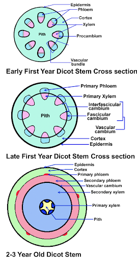
Early year dicot stem cross section and late first year dicot stem cross section.
Images by Dr. Thomas L. Rost, emeritus professor at UC-Davis.
Let’s start with the vascular cambium.

The three drawings on the right show a cross section of a stem for an imaginary woody dicot. The top drawing illustrates the stem early in the first year of growth, and shows the vascular cylinders arranged in a ring around the stem. The phloem is oriented to the outside, the xylem to the inside. A thin layer of parenchyma cells between the xylem and phloem has differentiated into the fascicular cambium (fascicular refers to bundles, in this case, cambium in the vascular bundles). The fascicular cambium is meristematic and can divide to produce new phloem toward the outside and new xylem to the inside. The new xylem and phloem produced by the cambium are called 2o (secondary) xylem and 2o phloem. Recall that the original xylem and phloem that differentiated from the apical meristem’s derivative cells are called the 1o (primary) xylem and 1o phloem.

The middle drawing is of the same stem later in the year. The cortex (cortical) parenchyma cells that lay between the vascular cylinders directly in line with the fascicular cambium begin to differentiate into a type of cambium called interfascicular cambium (cambium between the bundles). This is symbolized by the line connecting the vascular cylinders. This cambium is also meristematic, and produces 2o xylem and 2o phloem.

The cross section on the bottom illustrates the stem in its second or third year of growth, when there is a noticeable buildup of 2o xylem and 2o phloem with remnants of 1o xylem and 1o phloem.

In summary, the vascular cambium is a lateral meristem formed by differentiation of parenchyma cells located between the primary xylem and phloem into fascicular cambium, followed by differentiation of cortical parenchyma between the vascular cylinders into interfascicular cambium. After a few years of secondary growth, fascicular and interfascicular cambium can no longer be distinguished, and it is all simply known as vascular cambium. This layer of cambium runs vertically (assuming that the stem is oriented vertically) and parallel to the surface of the woody stem.

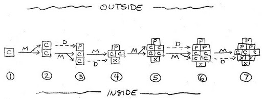
The illustration below shows how the cambium divides to produce 2o xylem and 2o phloem, with the outside of the stem toward the top of the page. Frame #1 shows a single cambium cell (C). This cell divides mitotically (M) to form two cambium daughter cells (Frame #2). Frame #3 shows that the cambium cell on the top differentiates (D) into a phloem cell (P-toward the outside) and the other cambium cell divides mitotically (M).

Cell division and differentiation from cambium to xylem/phloem
Cell division and differentiation from cambium to xylem/phloem. Tom Michaels
This type of cell division, in which new cells are formed either to the outside or inside, and the cell wall that separates the two new cells is parallel to the outside of the stem, is called periclinal division. Periclinal division by the cambium makes new cells that add girth to the plant. The cells that are added subsequently differentiate into xylem and phloem depending on their location to the outside or inside of the cambium. The meristem needs to divide periclinally to add girth to the plant stem.

In Frame #4, pay particular attention to a different type of cell division, where the cambium cell has divided so that the wall between the two cells is perpendicular to the outside of the stem. This is called anticlinal division. The meristem occasionally needs to divide anticlinally because as the stem is growing in girth, the diameter of the ring of vascular cambium must expand to keep up, or it will split into pieces and no longer form a continuous ring around the stem. Frame #4 also shows that the cambium cell to the inside has differentiated into xylem (X). In Frame #5, the two cambial cells that formed from anticlinal division now each divide periclinally.
 

Attachments

  • 1732250566221.png
    1732250566221.png
    5.7 MB · Views: 9

acespicoli

Well-known member
Mentor
1732251213775.png

1732251278862.png

Cross-sections of the top part of a hemp stem, stained with Calcofluor White. (A and B) Primary phloem fibres at a distance of 10 mm from the SAM. The fibre bundles are being formed in the inner part of the cortex; parenchyma cells (p) and laticifers (l) are present between the fibres and the procambium. (C and D) Primary phloem fibres at a distance of 80 mm from the SAM. Bundles of primary phloem fibres (pf) are already formed meaning that the intrusive growth has ceased. Fibres located at the bundle side closest to the stem periphery are the first to start secondary cell wall deposition. e, epidermis; col, collenchyma; pc, procambium; pf*, primary phloem fibres with thickened cell wall; tr, trichome. Bar: A and C ¼ 50 mm, B and D ¼ 20 mm.​

1732251327796.png

1732251374197.png

Explains why the most mature growth resists rooting :thinking:
Rotating mother plants...re-read this paper

 
Last edited:

acespicoli

Well-known member
Mentor
1732251572152.png

1732251671986.png

T1 - Non-photoperiodic transition of female cannabis seedlings from juvenile to adult reproductive stage
DO - 10.1007/s00497-022-00449-0
JO - Plant Reproduction
 

H e d g e

Well-known member
I often remove the leaf tips because I root them in a little bit of compost at the bottom of a small pot covered with clear plastic and they don’t fit with the leaves left whole.
Anyway, according to this Canadian research I shouldn’t..
866C6F5F-FACC-4467-BB1E-B79BB12C89D3.png
 

acespicoli

Well-known member
Mentor
I often remove the leaf tips because I root them in a little bit of compost at the bottom of a small pot covered with clear plastic and they don’t fit with the leaves left whole.
Anyway, according to this Canadian research I shouldn’t..
View attachment 19104216
They are usually citing the thought that the plants cannabilize the large fan leaves a nutrient reservoir for growth of roots. Always thought the success rate was better with snipped off shortened leaves. Enough to prevent drooping wilting and rot...we need to deep dive that and see what their idea of leaf removal involves.
 

Latest posts

Latest posts

Top